Multimodal AI platform can accurately diagnose and stage thyroid cancer via ultrasound images
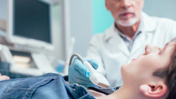
Using multiple artificial intelligence methods simultaneously, researchers were able to develop a platform that can accurately diagnose and stage thyroid cancer based off routine ultrasound images.
The results of the study are set to be presented at the 2022 Multidisciplinary Head and Neck Cancers Symposium by study author Annie Chan, MD, with the department of radiation oncology at Massachusetts General Hospital.
“We have developed an artificial intelligence platform that would examine ultrasound images and predict with high accuracy whether a potentially problematic thyroid nodule is, in fact, cancerous,” Chan said. “If it is cancerous, we can further predict the tumor stage, the nodal stage and the presence or absence of BRAF mutation.”
To develop their platform, researchers used a combination of four different AI methods: radiomics, topological data analysis, deep learning and machine learning. Each method has its own unique advantages for diagnosing diseases and predicting outcomes.
“By integrating different AI methods, we were able to capture more data while minimizing noise. This allows us to achieve a high level of accuracy in making predictions,” Chan explained.
This particular AI platform was trained using ultrasound images of 511 thyroid nodules from 464 patients. The images were divided into an internal training and validation set and an external validation set.
At 98.7%, the multimodal platform achieved significantly higher accuracy for diagnosing thyroid cancer compared to the individual AI methods using the internal dataset. For the external dataset, the platform again earned high marks, diagnosing nodule malignancy with 93% accuracy.
Additionally, the experts note the accuracy of the model’s ability to predict BRAF mutations. Employing a fivefold cross validation resulted in an accuracy of 96% in forecasting the genetic alteration.
The authors of the study suggest that their approach is low-cost and reproducible and can be utilized to develop personalized treatment for patients with thyroid cancer.
"If caught early, this disease is highly treatable, and patients generally can expect to live a long time after treatment," Dr. Chan suggested.
The results of this study are set to be presented on Feb. 25 at the 2022 Multidisciplinary Head and Neck Cancers Symposium, which is taking place in Phoenix, Arizona.
More on thyroid imaging and thyroid cancer:
FDA greenlights AI software that helps diagnose breast and thyroid cancers
Follow-up ultrasound for incidental thyroid nodules on CT not always cost-effective
'Highly significant' MRI findings link hyperthyroidism to structural brain abnormalities
ACR TI-RADS recommends 25%-50% fewer biopsies than other commonly used thyroid systems

