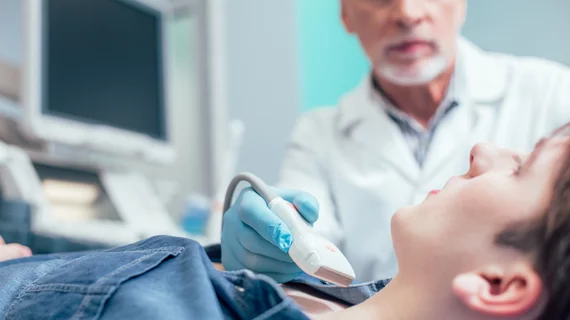
DL networks augment radiologist performance for thyroid cancer detection
Thyroid nodules are quite common, which makes differentiating the benign from the malignant masses an important diagnostic process—one that research suggests could benefit from the use of convolutional neural networks.
A new paper published in the European Journal of Radiology highlights how different deep learning models fared when classifying thyroid nodules on ultrasound images. After being presented with more than 15,000 images, each DL network yielded results comparable to that of 4 seasoned radiologists. This led the authors of the study to suggest that various models could play a complimentary role in the detection of thyroid cancer.
“The recent emergence of deep learning (DL) applications in US imaging has provided tremendous potential for improving the diagnostic performance in various organs, including differentiating hepatic fibrosis, metastatic cervical lymph nodes, malignant breast lesions, and thyroid nodules,” corresponding author Yangsean Choi, Clinical Assistant Professor, Department of Radiology at Seoul St. Mary’s Hospital College of Medicine, and co-authors explained.
Researchers used images from 7,321 patients who underwent ultrasound-guided fine-needle aspiration between January 2010 and March 2020 to train 3 different deep learning models —VGG16, VGG19, and ResNet—to distinguish benign thyroid nodules from malignant ones. The models’ performances were then compared to that of 4 radiologists.
The DL models performed better than the radiologists on the internal test set, but not the external test set. However, the numbers were still comparable. The highest performing model, VGG16, recorded an AUC of 0.86, sensitivity of 91.8% and specificity of 73.2% on the internal set compared to an AUC of 0.83, sensitivity of 78.6% and specificity of 76.8% on the external test set.
“One of the strengths of our study is the large number of pathologically confirmed thyroid nodules collected over a long period of time and the application of three well-known DL algorithms for image classification in search of optimal performance,” the authors wrote. “Furthermore, performance was directly compared with those of four radiologists, which allowed the approximation of DL models relative to practicing radiologists. Our results demonstrated an efficient pipeline for the detection and classification of thyroid nodules on US images.”
The experts went on to suggest that deep learning models could positively augment radiologists’ performance when identifying thyroid cancer, and that future studies should focus on models’ utility of classifying subtypes of thyroid malignancies.
Related ultrasound imaging content:
AI model could open doors for greater access to obstetric ultrasound
Experts suggest new follow-up imaging protocols for vaccine-related lymphadenopathy
O-RADS externally validated for differentiating between benign and malignant ovarian lesions

