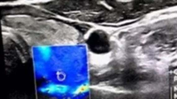
Shear wave elastography assessment improves TI-RADS scoring
Evaluating the vascularity and elastography in suspicious Thyroid Imaging Reporting and Data System (TI-RADS) categories was recently proven an effective strategy for more accurately assessing thyroid nodules for malignancy.
That’s according to an award-winning scientific online poster presented this week during the American Roentgen Ray Society’s annual meeting being held in Honolulu, Hawaii. TI-RADS scores derived from conventional ultrasound exams are the standard of care for evaluating thyroid nodules, but researchers hypothesized that the addition of shear wave elastography—an ultrasound technique that analyzes tissue stiffness—could offer radiologists a more in-depth look at the nodules.
For the work, Leila Aghaghazvini, MD, from the Department of Radiology at Shariati Hospital and Iran’s University of Medical Sciences in Tehran, and colleagues assessed 200 thyroid nodules. Conventional ultrasound was used for TI-RADS scoring and to obtain anatomical details, followed by the addition of shear wave elastography.
The team used a 7.5 MHz probe to evaluate vascularity characteristics and resistive index (RI). Quantitative elastography was assessed by way of color mapping and determining mean and maximum velocities in shear-wave mode. Histopathology and follow-up imaging were used to confirm diagnoses.
Out of the 200 nodules included in the study, 27 were found to be malignant—14 were papillary thyroid carcinoma, 11 were follicular thyroid carcinoma and 2 were medullary thyroid cancer. TI-RADS assessments from conventional ultrasound yielded an AUC of .76. Out of 37 TI-RADS category 4 nodules, just 15 were deemed malignant, while 12 out of 57 T-IRADS category 5 nodules were cancerous.
Adding SWE to the equation improved these metrics. In TI-RADS 4 only, Doppler grade AUC, RI, color map elastography grade, SWE maximum velocity, and mean SWE velocity for diagnosing malignancy were 0.8, 0.93, 0.89, 0.86, and 0.82, respectively. And for TI-RADS 5, those measures were 0.8, 0.96, 0.85, 0.96, and 0.97.
For additional insight into the findings, click here.

