Computed Tomography

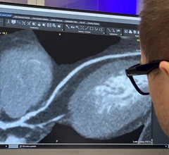
Cardiac computed tomography (CT) has become a primary cardiovascular imaging modality in the past 20 years, and was recommended as a 1A recommendation in the 2021 chest pain assessment guidelines. CT calcium scoring has became a primary risk assessment for coronary artery disease and whether patients should be on statins. Coronary CT angiography (CCTA) is used to for anatomical assessment of the arteries for plaque burden and to identify areas of blockage that may cause ischemia and heart attacks. Additional use of contrast CT perfusion or fractional flow reserve CT (FFR-CT) can offer physiological information on the function of the heart. CT plays a primary role in structural heart assessments for heart valves, repair of congenital defects and left atrial appendage occlusion (LAAO) for both pre-procedure planning and procedural guidance. Find more news on general radiology CT use.

AI cardiology heart artificial intelligence deep learning

FDA approves new AI-powered imaging software from Nanox subsidiary

According to Nanox.AI, previously known as Zebra Medical Vision, the updated software helps "bridge the divide between radiology and cardiology."

Images coronary calcium imaging by optical coherence tomography (OCT) and high-definition intravascular ultrasound (IVUS).

OCT-guided PCI improves survival after life-threatening heart attacks

Researchers explored data from nearly 4,000 STEMI patients, noting that optical coherence tomography may be a valuable first step of any treatment strategy. 

Ron Blankstein, MD, FACC, FASNC, MSCCT, FASPC, associate director, cardiovascular imaging program, director, cardiac computed tomography, Brigham and Women’s Hospital, and a professor of medicine and radiology, Harvard Medical School, explains a few of the recent advancements in cardiac MRI, nuclear imaging and computed tomography.

Exploring major breakthroughs in cardiac imaging

Ron Blankstein, MD, director of cardiac computed tomography for Brigham and Women’s Hospital, details recent developments in cardiac MRI, nuclear imaging and CT.

CMS may double Medicare payments for cardiac CT

The agency is gathering feedback on a proposal to double the amount hospitals are paid for coronary computed tomography angiography, with the comment period ending Sept. 9.

HeartFlow introduced its next generation artificial intelligence (AI) Plaque Analysis with an interactive experience at SCCT 2024. It shows a 3D plaque model and analysis by territory across calcified, non-calcified and low-attenuation plaques. This includes viewing cross-sectional, color-coded images of each plaque type where it was quantified along the vessel. #SCCT #SCCT24 #SCCT2024

Updated HeartFlow technology offers interactive features, full integration with FFR-CT 

HeartFlow is using SCCT 2024 to introduce the world to its updated Plaque Analysis platform. “Accurately diagnosing a patient’s risk for coronary artery disease is critical for determining the best treatment," Chief Medical Officer Campbell Rogers, MD, explained.

Researchers with University College London and the European Synchrotron Radiation Facility (ESRF) have used a new X-ray technique, hierarchical phase-contrast tomography (HiP-CT), to capture images of the human heart in unprecedented detail. The group shared its images, as well as a full analysis, in Radiology.[1]

New imaging technique captures human heart with 'unprecedented detail'

The new images were captured at the European Synchrotron Radiation Facility using hierarchical phase-contrast tomography. One specialist called them "Google Earth for the human heart." 

SCCT aims to provide common language for CCTA use with updated guidance

The Society of Cardiovascular Computed Tomography has developed a new expert consensus document designed to get physicians on the same page when discussing this growing technology. 

The rapid rise of artificial intelligence (AI) has helped cardiologists, radiologists, nurses and other healthcare providers embrace precision medicine in a way that ensures more heart patients are receiving personalized care.

AI uses imaging results to ID high-risk TAVR patients with speed, accuracy

Researchers developed an advanced AI model capable of extracting measurements from unprocessed CT images in seconds. It then uses those data to evaluate the patient's mortality risk if they underwent TAVR.

Around the web

GE HealthCare designed the new-look Revolution Vibe CT scanner to help hospitals and health systems embrace CCTA and improve overall efficiency.

Clinicians have been using HeartSee to diagnose and treat coronary artery disease since the technology first debuted back in 2018. These latest updates, set to roll out to existing users, are designed to improve diagnostic performance and user access.

The cardiac technologies clinicians use for CVD evaluations have changed significantly in recent years, according to a new analysis of CMS data. While some modalities are on the rise, others are being utilized much less than ever before.