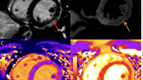
New cardiac MRI analysis offers updated insight into long-term impact of vaccine-related myocarditis
Follow-up imaging of patients who experienced myocarditis after receiving their COVID vaccine offers additional insight into how these individuals have fared in the months following their initial recovery.
A paper published in Radiology last February concluded that patients who had been diagnosed with acute myocarditis post-inoculation tended to have only mild cases, for which they recovered from quickly. And compared to patients who developed myocarditis as a result of contracting COVID-19, the group with vaccine-related inflammation visualized on cardiac MRI was reported to have less severe cases that resulted in less functional impairment.
“We previously reported cardiac MRI findings in a cohort of patients with COVID-19 vaccine associated myocarditis. The majority of patients had mild imaging abnormalities at the time of acute symptoms,” corresponding author Kate Hanneman, MD, MPH, cardiac radiologist at Toronto General Hospital, University Health Network, and co-authors discussed in a letter to the editor. “However, little is known about later evolution of the cardiac MRI abnormalities.”
The follow-up analysis consisted of 13 patients who had been previously diagnosed with vaccine-related myocarditis. Upon initial imaging for the acute condition, all patients presented with chest pain, 46% of whom required hospitalization. Patients underwent a follow-up cardiac MRI 74 to 237 days after vaccination. At that time, none of the patients were symptomatic, nor had any experienced an adverse cardiac event. Troponin levels were reported to be normal upon follow-up as well.
The most recent cardiac MRI scans revealed that myocardial edema observed on previous imaging had resolved in all patients, while left ventricular function had also normalized. Late gadolinium enhancement (LGE) had resolved completely for 23% of patients, decreased for 62% and remained negative in 15%. This, the authors explained, could reflect myocardial fibrosis.
“These findings are consistent with the typical rapid decrease in myocardial inflammation in other causes of myocarditis,” the authors wrote. “In conjunction with lack of any adverse events at 5-month follow-up, this raises the likelihood that vaccine-associated myocarditis might have a favorable prognosis despite the persistence of minimal LGE.”
The entire report, Cardiac MRI and Clinical Follow-up in COVID-19 Vaccine–associated Myocarditis, can be viewed in Radiology.
Related cardiac imaging content:
Cardiac MRI scans offer new insight into COVID vaccine-related myocarditis
Study urges radiologists to report CAC findings on all chest CTs, regardless of clinical indication
Risks of stroke and heart attack increase with larger thoracic aortic diameter, research shows

