Magnetic Resonance Imaging

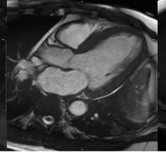
Cardiac magnetic resonance imaging MRI is used as both a functional and anatomical cardiac imaging test. It offers excellent soft tissue detail and the ability to quantify cardiac function. MRI scans can be performed with or without gadolinium contrast depending on what information is needed. Unlike computed tomography (CT), MRI does not use X-ray radiation, but patients with metal implants may have contraindications for MRI use because MR will heat up most metal objects. MRI exams usually take much longer than CT scans. How does MRI work? MR creates images by using powerful magnets to polarize hydrogen atoms in water (the body is made of of more than 80% water) so they face in one direction. A radiofrequency pulse is then used to ping these atoms, causing them to wobble, or resonate. The MRI coils detect this and computers can assemble images from the signals. Basic MRI scans will focus on the resonance of fat and water in two different sequences, which highlight and contrast different features in the anatomy.
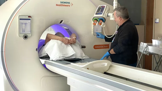
GE HealthCare SIGNA Sprint MRI

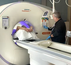
GE HealthCare debuts new wide-bore MRI scanner built with cardiology, oncology in mind

The new 1.5T MRI scanner includes a wide bore and key AI features designed to boost the patient experience.

Imricor Medical Systems ventricular ablation

Cardiologists make history, perform world’s first ventricular ablation guided by real-time MRI

“MRI images provide a better view of the anatomy of the heart and the instruments used for treatment, but also of the changes made to the treated cardiac tissue,” one cardiologist explained. “Unlike X-ray images, the entire area surrounding the heart can be seen, including the blood vessels and valves."

Key trends in diagnostic heart testing: CT on the rise as some traditional techniques fall out of favor

The cardiac technologies clinicians use for CVD evaluations have changed significantly in recent years, according to a new analysis of CMS data. While some modalities are on the rise, others are being utilized much less than ever before.

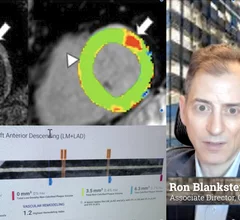
MRI technique analyzes composition of fat surrounding heart.

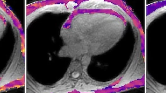
New MRI technique spots signs of heart disease before symptoms emerge

Researchers with the University of Virginia Arlington are working to develop a new way of analyzing fat content around the heart. 

artificial intelligence AI heart cardiology

Philips, Mayo Clinic using AI to improve cardiac MRI technology

The two companies aim to improve patient access to high-quality MRI scans by combining their artificial intelligence capabilities.

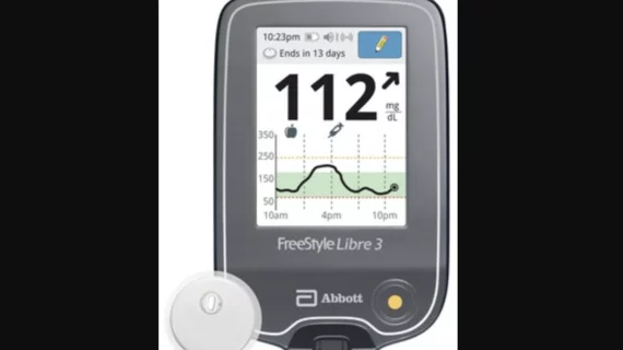
Abbott’s FreeStyle Libre 3 integrated continuous glucose monitoring (iCGM) system

Abbott CGM sensors can now be worn during common imaging procedures

Sensors from the FreeStyle Libre 2 and Libre 3 continuous glucose monitoring systems can now be worn during X-rays, CT scans and MRI scans. The news represents a shift in policy from the FDA, one that came after the agency reviewed extensive testing data.

The American Society of Echocardiography (ASE) has shared new recommendations for performing medical imaging exams on heart failure patients with surgically implanted left ventricular assist devices (LVADs) and temporary mechanical circulatory support (TMCS) devices. The guideline, published in full in the Journal of the American Society of Echocardiography, represents ASE’s first update on the topic since 2015.[1]

ASE shares new guideline on imaging patients with implanted devices

The new guideline details the best imaging strategies for a variety of clinical scenarios. 

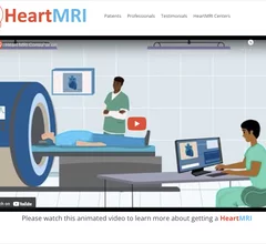
The Society of Cardiovascular Magnetic Resonance (SCMR) recently launched HeartMRI.org, which provides includes a general information and videos for patients and referring physicians.

SCMR launches cardiac MRI educational video to assist referring physicians 

The Society of Cardiovascular Magnetic Resonance launched a new website aimed at patients and referring physicians that includes pamphlets and videos on cardiac MRI. 

Around the web

These findings present additional evidence that invasive imaging tests are not necessarily more effective when it comes to evaluating patients for chest pain.

Unlike other UEA options, GE HealthCare's Optison does not contain polyethylene glycol. The FDA approved its use for adult patients back in 1997.

The new 1.5T MRI scanner includes a wide bore and key AI features designed to boost the patient experience.