Cardiac Imaging

While cardiac ultrasound is the widely used imaging modality for heart assessments, computed tomography (CT), magnetic resonance imaging (MRI) and nuclear imaging are also used and are often complimentary, each offering specific details about the heart other modalities cannot. For this reason the clinical question being asked often determines the imaging test that will be used.

Thumbnail

Cardiologists ID signs of widespread heart disease in ancient mummies

Researchers evaluated vascular samples from more than 200 mummies, presenting their findings in European Heart Journal.

fasting before tavr or ablation

Fasting before coronary angiography, PCI may not be necessary

Relaxing fasting restrictions before interventional coronary procedures improves patient comfort—and there does not appear to be much of a downside. 

medical imaging carbon emissions climate change

PET is an energy hog, but relatively rare utilization lessens its carbon footprint. MRI and CT have no such ‘out’

Medical imaging machinery uses a lot of power, and the extent to which it quickens the pace of global warming is becoming clearer.

AI-based CAD assessments dramatically improve vascular surgery outcomes

PAD patients evaluated with HeartFlow's noninvasive FFRCT Analysis technology prior to surgery experienced much better outcomes, including a 63% lower risk of all-cause mortality after five years. 

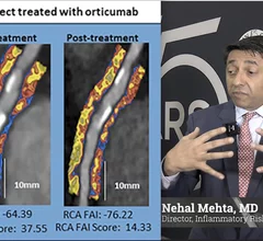
Interview with Nehal Mehta, MD, Penn Medicine, who explains how coronary inflammation can be seen using AI on cardiac CT scans to better risk stratify patients and begin preventive drug therapy.

AI helps cardiologists track new drug's effect on inflammation

The combination of AI and CT helped Nehal Mehta, MD, and colleagues track the performance of a new drug designed to target coronary inflammation. 

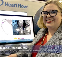
Sarah Jane Rinehart, MD, director of cardiac imaging, Charleston Area Medical Center, Charleston West Virginia, as been using the FDA-cleared RoadMap artificial intelligence algorithm from HeartFlow in studies and in clinical used since it was cleared and said it helps cardiologists in several ways. #ACC #ACC24 #ACC2024 #Heartflow #AIhealth

AI improves CT assessments, boosts care for real-world heart patients

Automated AI-generated measurements combined with annotated CT images can improve treatment planning and help referring physicians and patients better understand their disease, explained Sarah Jane Rinehart, MD, director of cardiac imaging with Charleston Area Medical Center.

Advanced artificial intelligence (AI) models can evaluate cardiovascular risk in routine chest CT scans without contrast, according to new research published in Nature Communications.[1] In fact, the authors noted, the AI approach may be more effective at identifying issues than relying on guidance from radiologists.

AI predicts cardiovascular risk during CT scans—no invasive tests or contrast required

Two advanced algorithms—one for CAC scores and another for segmenting cardiac chamber volumes—outperformed radiologists when assessing low-dose chest CT scans. 

Thumbnail

CT procedure shown to reduce need for invasive cardiac testing

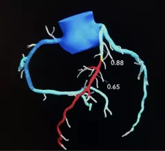
In a study, CT-FFR was shown to reduce the need for invasive tests to measure coronary artery blockage from 74.5% to 25.5%.

Around the web

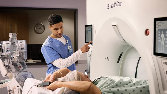
GE HealthCare designed the new-look Revolution Vibe CT scanner to help hospitals and health systems embrace CCTA and improve overall efficiency.

Clinicians have been using HeartSee to diagnose and treat coronary artery disease since the technology first debuted back in 2018. These latest updates, set to roll out to existing users, are designed to improve diagnostic performance and user access.

The cardiac technologies clinicians use for CVD evaluations have changed significantly in recent years, according to a new analysis of CMS data. While some modalities are on the rise, others are being utilized much less than ever before.