Cardiac Imaging

While cardiac ultrasound is the widely used imaging modality for heart assessments, computed tomography (CT), magnetic resonance imaging (MRI) and nuclear imaging are also used and are often complimentary, each offering specific details about the heart other modalities cannot. For this reason the clinical question being asked often determines the imaging test that will be used.

doctor examines patient data on their tablet

FDA sees potential in new PET imaging agent for cardiac amyloidosis

Early evidence suggests a new PET imaging agent from California-based Attralus can help evaluate all varieties of systemic cardiac amyloidosis. It has now been granted the FDA's breakthrough therapy designation. 

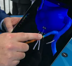
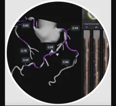
HeartFlow introduced its next generation artificial intelligence (AI) Plaque Analysis with an interactive experience at SCCT 2024. It shows a 3D plaque model and analysis by territory across calcified, non-calcified and low-attenuation plaques. This includes viewing cross-sectional, color-coded images of each plaque type where it was quantified along the vessel. #SCCT #SCCT24 #SCCT2024

Updated HeartFlow technology offers interactive features, full integration with FFR-CT 

HeartFlow is using SCCT 2024 to introduce the world to its updated Plaque Analysis platform. “Accurately diagnosing a patient’s risk for coronary artery disease is critical for determining the best treatment," Chief Medical Officer Campbell Rogers, MD, explained.

Researchers with University College London and the European Synchrotron Radiation Facility (ESRF) have used a new X-ray technique, hierarchical phase-contrast tomography (HiP-CT), to capture images of the human heart in unprecedented detail. The group shared its images, as well as a full analysis, in Radiology.[1]

New imaging technique captures human heart with 'unprecedented detail'

The new images were captured at the European Synchrotron Radiation Facility using hierarchical phase-contrast tomography. One specialist called them "Google Earth for the human heart." 

SCCT aims to provide common language for CCTA use with updated guidance

The Society of Cardiovascular Computed Tomography has developed a new expert consensus document designed to get physicians on the same page when discussing this growing technology. 

Researchers have developed and validated a new artificial intelligence (AI) model capable of producing four-chamber cardiac MR (CMR) images in seconds, publishing their findings in European Radiology Experimental.

AI creates accurate 4D heart scans in seconds

New data out of England provides cardiologists and radiologists with another reminder of AI's potential to transform healthcare. 

ASE President Ted Abraham, MD, speaking at the ASE HCM forum in 2023. Photo by Dave Fornell

Cardiologist begins term as American Society of Echocardiography's new president

Cardiologist Theodore Abraham, MD, has been an ASE member for more than two decades. He is now the organization's 2024-2025 president. 

Stretching may help reduce painful orthopedic issues among echo and interventional lab staff

Orthopedic issues are the biggest work-related injury in cardiology, but this low cost intervention may help.

The rapid rise of artificial intelligence (AI) has helped cardiologists, radiologists, nurses and other healthcare providers embrace precision medicine in a way that ensures more heart patients are receiving personalized care.

AI uses imaging results to ID high-risk TAVR patients with speed, accuracy

Researchers developed an advanced AI model capable of extracting measurements from unprocessed CT images in seconds. It then uses those data to evaluate the patient's mortality risk if they underwent TAVR.

Around the web

GE HealthCare's flurpiridaz, the PET radiotracer that recently received FDA approval, offers several key benefits over SPECT. Jamshid Maddahi, MD, discussed the details in an exclusive interview. 

Ultrafast MCE could go on to become a go-to treatment option for obstructive coronary artery disease, according to the authors of a new first-in-human clinical study.

Elucid's PlaqueIQ was trained to turn CCTA images into interactive 3D reports that help physicians visualize the presence of atherosclerosis.

Trimed Popup
Trimed Popup