PACS

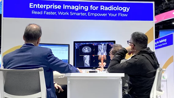
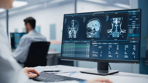
Picture archiving and communication systems (PACS) have replaced conventional radiographic films as the digital image-viewing hub over the past two decades and now serve as the primary communication bridge between radiologists, radiologic technologists and referring providers. PACS enables all authorized clinicians to access medical images and reports quickly, easily and from virtually any location. Some health systems have integrated PACS into the electronic medical record (EMR). Others have moved to enterprise image systems to replace radiology PACS and allow all departments to now store images and reports in one location for easier health system-wide access.

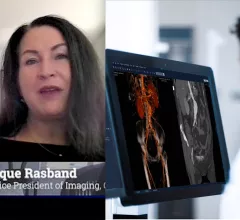
Monique Rasband from KLAS Research shares trends in PACS and radiology informatics.

VIDEO: 6 key trends in PACS and radiology informatics observed by KLAS

Monique Rasband, vice president of imaging, cardiology and oncology, KLAS Research, shares some of technology trends observed in radiology PACS and and imaging informatics since 2019.

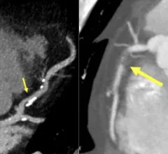
Examples of new plaque reporting in the CAD-RADS 2.0 document. Left, an example from CAD-RADS 2 / P2 plaque burden with mild coronary stenosis (25-49%). Right, example of a CAD-RADS 5/ P3, with a focal, non-calcified occlusion of the proximal RCA (arrow) and severe amount of plaque (P3). #CADRADS #YesCCT #CTA #CCTA

New CAD-RADS 2.0 reporting for coronary CTA offers patient management recommendations

The document includes updated classification to established a framework for stenosis, plaque burden and plaque modifiers, including assessment of CT-FFR or myocardial CT perfusion.

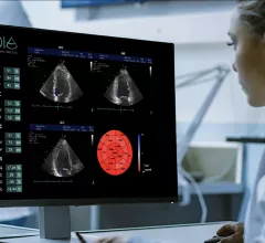
DiA Imaging Analysis, which specialized in developing the AI-based automated cardiac ultrasound solution LVivo Seamless. The technology is now integrated through partnerships with dozens of healthcare vendors, including ScImage, GE Healthcare, Philips Healthcare Konica Minolta and IBM Watson.

ScImage latest vendor to adopt DiA Imaging Analysis AI for echocardiography

Artificial intelligence vendor DiA has emerged as a key third-party provider of AI to larger imaging vendors.

Thumbnail

VIDEO: What to look for in radiology workflow orchestration software

Elizabeth Bergey, MD, a diagnostic radiologist at Quantum, chairman of Quantum’s Board of Directors, discusses some of the questions radiology practices should be asking when evaluating orchestration software.

Thumbnail

How to prevent cherry picking on radiology worklists

One of the problems in radiology today is the selective cherry picking of the easier, more desirable cases from the DICOM worklists and leaving the more complicated studies for other radiologists, which is one of the factors in radiologist burnout.

A radiologists reading station, image from Sectra

VIDEO: How to Prevent Radiologist Burnout

Elizabeth Bergey, MD, a diagnostic radiologist at Quantum, chairman of Quantum’s board of directors, explains some of the issues that cause burnout and how technology can help mitigate issues that cause radiologists to leave.

Thumbnail

Managing incidental radiologic findings: ACR-led initiative proposes several recommendations

Authors of the JACR paper noted the timeliness of the proposed measures, referring to CMS’ prioritization of measures of patient outcomes.

Elizabeth Bergey, MD, a diagnostic radiologist at Quantum, explained how the radiology group orchestrates its work lists for load balancing at the 2022 Radiology Business Management (RBMA) meeting.

VIDEO: Approaches to intelligent radiologist worklist orchestration

Elizabeth Bergey, MD, a diagnostic radiologist at Quantum, chairman of Quantum’s Board of Directors, explains how the radiology group orchestrates its work lists for load balancing using an AI-driven IT system for its work lists.

Around the web

The new technology shows early potential to make a significant impact on imaging workflows and patient care. 

Richard Heller III, MD, RSNA board member and senior VP of policy at Radiology Partners, offers an overview of policies in Congress that are directly impacting imaging.
 

The two companies aim to improve patient access to high-quality MRI scans by combining their artificial intelligence capabilities.