RSNA releases expert guidelines for reporting COVID-19 radiology findings
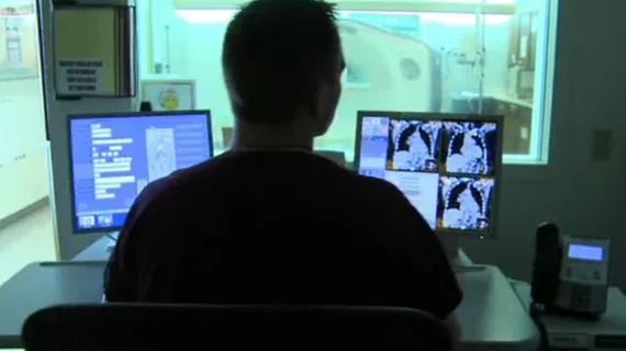
The Radiological Society of North America on Wednesday published a consensus statement developed by imaging experts across the U.S. to help radiologists identify chest CT findings of COVID-19 and reduce reporting variability.
A group of nine academic medical center leaders created the guidance, which has been endorsed by the American College of Radiology and the Society of Thoracic Radiology. Published March 25 in Radiology: Cardiothoracic Imaging, the document includes four categories for reporting CT imaging findings of possible COVID-19, including standardized language.
Editor of the journal and chief of the Cardiothoracic Imaging Division at UT Southwestern Medical Center in Dallas, Texas, Suhny Abbara MD, said the tool will improve communication between providers and help limit physicians’ anxiety when managing the overwhelming number of incoming cases.
“The goal of this expert consensus is to help radiologists recognize findings of COVID-19 pneumonia and aid their communication with other healthcare providers, assisting management of patients during this pandemic,” the authors wrote. "We believe it is important to provide radiologists and referring providers guidance and confidence in reporting these findings and a more consistent framework to improve clarity," Abbara said in an RSNA statement.
Most radiology societies are not currently recommending routine screening CT for diagnosing the new virus, but the volume of these exams performed for suspected cases has certainly increased, the authors noted. And during these tests, some radiologists may spot incidental findings suggestive of COVID-19 pneumonia, forcing them to decide whether or not to mention the virus as a possibility in their reporting.
Doing so can set off a number of events, including infection control measures and anxiety for providers and patients, the authors wrote. Similarly, teleradiology company vRad recently urged caution for citing the coronavirus in imaging reports, pointing to potential economic and workflow implications.
Abbara and colleagues propose using four categories for reporting CT findings: typical appearance, indeterminate appearance, atypical appearance and negative for pneumonia. Importantly, the authors noted that their document is merely a guide for reporting and that radiologists should work with their individual institutions to establish a tailored approach.
“The reporting language does not offer an exact likelihood for COVID-19 pneumonia, which depends on several factors including prevalence in a community, exposure, risk factors, and clinical presentation,” they went on to say. “Rather, the reporting language focuses on CT findings reported in the literature and the typicality of these features in COVID-19 pneumonia rather than other diseases.”
Clinical understanding and knowledge of the coronavirus is constantly evolving, the researchers noted, but radiologists should always adhere to the ACR Practice Parameters for Communication of Diagnostic Imaging Findings.
For more detailed guidelines, read the entire statement here.

