Nuclear Medicine

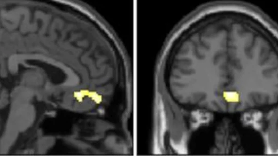
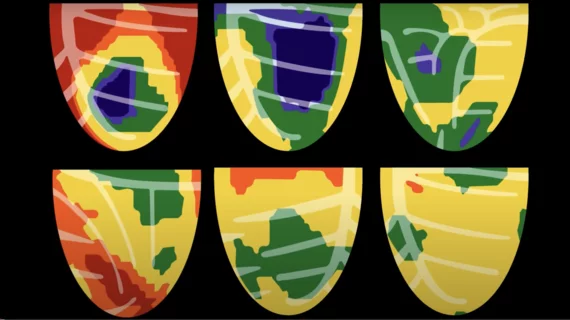
Nuclear medicine (also called molecular imaging) includes positron emission computed tomography (PET) and single photon emission computed tomography (SPECT) imaging. Nuclear imaging is achieved by injecting small amounts of radioactive material (radiopharmaceuticals) into patients before or during their scan. These can use sugars or chemical traits to bond to specific cells. The radioactive material is taken up by cells that consume the sugars. The radiation emitted from inside the body is detected by photon detectors outside the body. Computers take the data to assemble images of the radiation emissions. Nuclear images may appear fuzzy or ghostly rather than the sharper resolution from MRI and CT.  But, it provides metabolic information at a cellular level, showing if there are defects in the function of the heart, areas of very high metabolic activity associated with cancer cells, or areas of inflammation, data not available from other modalities. These noninvasive imaging exams are used to diagnose cancer, heart disease, Alzheimer’s and Parkinson’s disease, bone disorders and other disorders. 

UltraSPECT and ASNC partner to provide technologist travel awards to attend ASNC2014

UltraSPECT, provider of the most cost-effective solutions for meeting American Society of Nuclear Cardiology (ASNC) guidelines for low dose nuclear medicine imaging, announces today its support of nuclear medicine technologists via six new grants.

Disparities based on age, race persist in PET use for lung cancer patients

Demographic differences in the use of PET imaging among Medicare beneficiaries with non-small cell lung cancer have persisted since the modality’s approval by Medicare in 1998, according to a study published online Feb. 15 in Radiology.

Thumbnail

PET technique visualizes amyloid deposits in heart

PET with 11C-PIB provides a noninvasive method for visualizing amyloid deposits in the heart, according to a study published in the February issue of the Journal of Nuclear Medicine. The researchers suggest that 11C-PIB eventually may be used in the clinical setting as both a diagnostic tool and a treatment follow-up method.

ASNC Releases Dose-Reduction Guidelines for Nuclear Cardiology

Technological improvements in image acquisition and software processing in nuclear cardiology should allow physicians to shave patient imaging times dramatically or cut radiation doses fourfold, according to a new preferred practice statement from the American Society of Nuclear Cardiology (ASNC).

Around the web

GE HealthCare designed the new-look Revolution Vibe CT scanner to help hospitals and health systems embrace CCTA and improve overall efficiency.

Clinicians have been using HeartSee to diagnose and treat coronary artery disease since the technology first debuted back in 2018. These latest updates, set to roll out to existing users, are designed to improve diagnostic performance and user access.

The cardiac technologies clinicians use for CVD evaluations have changed significantly in recent years, according to a new analysis of CMS data. While some modalities are on the rise, others are being utilized much less than ever before.