Deep learning triages chest X-rays with tuberculosis, rivaling radiologists and pulmonary experts
A new deep learning model rivaled radiologists’ accuracy at assessing tuberculosis on chest X-rays. The tool may prove particularly useful for triaging patients in low-resource settings.
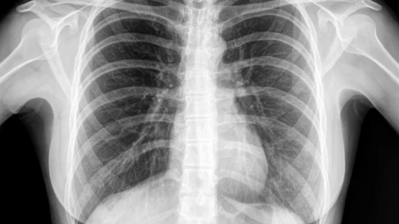
Chest radiography is among the top choices for detecting and diagnosing tuberculosis—one of the top 10 causes of death worldwide, researchers explained Tuesday in Radiology. Yet countries with higher rates of TB also have fewer experts to combat the problem.
With this in mind, South Korean providers developed a deep learning model to identify active pulmonary tuberculosis on chest images. They trained their tool using more than 6,500 radiographs taken from patients with active tuberculosis and those who had recovered.
Overall, the model performed well, distinguishing images with TB from healthy cases. It also beat out pulmonologists and performed similarly to radiologists, the authors noted.
“The network may help radiologically triage patients with active tuberculosis by excluding healed tuberculosis in high-burden countries and may assist in monitoring the activity of mycobacterial diseases that require long-term treatment,” Seowoo Lee, of Seoul National University Hospital’s Department of Radiology, and co-authors wrote in the study.
The neural network is based on 6,654 radiographs, including X-rays from 3,327 with pulmonary tuberculosis and 3,182 normal chest X-rays.
The team tested their model on two datasets: Set 1 included 89 patients and 148 radiographs, and set 2 utilized randomized controlled data from 366 patients and more than 3,700 images.
Two pulmonologists and two thoracic rads were blinded to disease activity info while they independently analyzed images from both datasets.
For test set 1, the platform notched a higher area under the receiver operating characteristic curve score (0.83) than one pulmonologist (0.69) and similar numbers as the radiologists (0.79-0.80). In set 2, the model reached a 0.84 AUC, higher than the pulmonary expert (0.71) and on par with the radiologists (0.79 and 0.80).
With further testing and validation, a tool like the one presented here could make a huge difference for many lives, the authors explained.
“If a deep learning network can accurately differentiate active tuberculosis from healed tuberculosis on radiographs, then this will provide more efficient triage for testing in limited-resource settings by decreasing unnecessary testing in individuals less likely to have active tuberculosis,” Lee et al. concluded.

