
Mycoplasma pneumonia: Experts highlight imaging findings linked to the pediatric diagnosis
Following a surge of mycoplasma pneumoniae pneumonia (MPP) in 2024, experts have issued new guidance to help providers quickly identify and treat the condition, with imaging playing a prominent role in the process.
MPP is a bacterial pathogen that causes pneumonia in children. The Centers for Disease Control and Prevention (CDC) estimates that MPP impacts around 2 million people annually. However, in 2023 and 2024, there was a noticeable uptick, with cases more than doubling in some age groups.
Depending on severity, the presentation of MPP can vary in children, making its diagnosis tricky in some cases. What’s more, many of the symptoms of MPP overlap with other respiratory conditions. As such, there is a need for standardized clinical criteria to accurately diagnose MPP early before symptoms become difficult to manage.
To address this, a group of multidisciplinary experts with the Chinese Medical Association collaborated to create a set of guidelines providers can use to identify MPP.
“Despite various laboratory diagnostic methods, the diagnostic criteria remain inconsistent, potentially leading to missed or overdiagnosis,” Professor Baoping Xu, from the Department of Respiratory Medicine at Beijing Children’s Hospital, and colleagues noted in the journal Pediatric Investigation. “The incidence of severe and refractory cases of MPP is increasing, and there are issues with nonstandard treatment in clinical practice.”
In addition to signaling preference for Polymerase chain reaction (PCR) testing and antibody detection analyses, the team also highlighted the importance of imaging in identifying true cases of MPP.
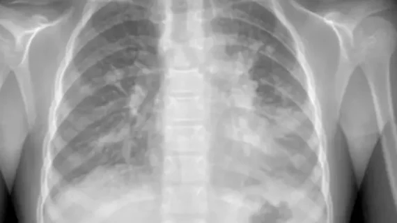
Chest X-rays are considered the gold standard for differentiating MPP from other pneumonias. However, the presentation of MPP on imaging can differ. To address these varying imaging manifestations, experts issued the following guidance for distinguishing MPP on radiographs:
During the acute phase of MPP, lobar or segmental consolidation and pleural effusion are common findings on chest X-rays, especially for children ages 6 and older.
Some patients also show lobular lesions, interstitial infiltration, emphysema and atelectasis.
Though beneficial, changes on imaging alone should not be used to determine MPP positivity. However, when the aforementioned imaging manifestations are present in children 6 and older, MPP should be considered.
In more severe cases, chest CT may be necessary to analyze inflammation.
"Accurate and timely diagnosis is crucial in preventing unnecessary antibiotic use and ensuring appropriate treatment," the group wrote. "By combining Polymerase chain reaction testing, antibody detection and imaging, we can improve diagnostic precision and reduce misdiagnosis."
The new guidelines also address different treatment strategies, taking into consideration the growing prevalence of antibiotic resistance among the population.
The full list of recommendations can be found here.

In addition to her background in journalism, Hannah also has patient-facing experience in clinical settings, having spent more than 12 years working as a registered rad tech. She began covering the medical imaging industry for Innovate Healthcare in 2021.