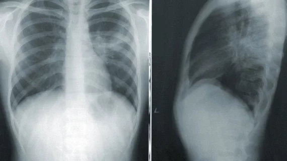
Chest x-ray can’t top lab testing, CT for COVID-19 but remains valuable
Some countries have turned to chest x-ray as a first-line tool to triage patients with the coronavirus, due to limited CT availability and long lab-testing turnaround times. But new research suggests the modality may not be up to snuff.
A group of experts based in Hong Kong compared the diagnostic ability of chest x-ray to that of real-time reverse transcription polymerase-chain-reaction testing in more than 60 patients with COVID-19. The results, published Friday in Radiology, suggest CXR yielded a sensitivity of 69% compared to 91% for lab testing.
Lead author Ho Yuen Frank Wong, with Queen Mary Hospital’s Department of Radiology, and colleagues also reported that the most common CT findings of the new virus were just as apparent on x-ray exams. Some prior research has suggested the opposite.
“As the COVID-19 pandemic threatens to overwhelm healthcare systems worldwide, CXR may be considered as a tool for identifying COVID-19, but is less sensitive than CT,” Wong et al. wrote.
Radiologists should keep this in mind, but some countries such as the U.K.—with limited imaging resources—have been forced to deploy chest x-ray to avoid overwhelming CT exam areas. And that may be okay, in some instances, the authors noted.
“Although further studies are needed, in the scenario where there is high clinical suspicion of COVID-19 it is conceivable that a positive CXR may obviate the need for a CT, thus reducing burden on CT units in this pandemic,” Wong and colleagues wrote.
The Hong Kong team retrospectively looked at 64 patients who were admitted to four area hospitals between January and March. Two expert radiologists analyzed each x-ray, looking at consolidation, ground-glass opacity, location and pleural fluid, and assigned a severity index to each lung.
Of the total group, 91% had a positive initial RT-PCR test; 69% showed abnormalities on baseline CXR and 59% had both. A total of six patients showed x-ray abnormalities prior to testing positive for the new virus via lab testing.
Wong and colleagues also pointed out that some COVID-19 patients can test positive on RT-PCR after they’re discharged, leading them to suggest imaging as a complementary tool to track disease progression, for now.
“With the situation further confounded by reports of COVID-19 patients testing positive on RT-PCR again after discharge, our opinion is that imaging should be used as an adjunct to clinical parameters in monitoring of disease course, until further evidence is available,” they concluded.

