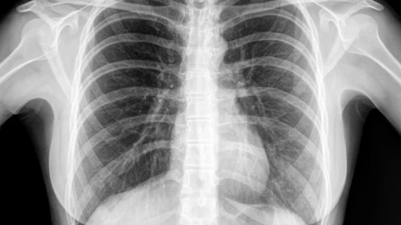
Deep learning model accurately detects COVID-19 on chest X-ray images
A deep learning model known as ResNet18 accurately classified over 99% of chest X-rays into three categories—COVID-19, other viral pneumonia, or normal health, according to a new study in Computational Intelligence.[1]
The results demonstrate the potential of such programs to help clinicians trying to detect COVID-19 in pneumonia patients, since it can often be difficult to distinguish in radiographic images, according to study author Onur Sevli, MD, of Budur Mehmet Akif Ersoy University in Turkey.
“The fact that different pathogens other than COVID-19 also cause pneumonia and the radiographic images of all are similar make it difficult to detect the source of the disease,” Sevli wrote. “This result reveals that the conducted study can play the role of a successful decision support system for experts.”
The study tested five existing models, each using a deep learning technique known as a neural network, on a dataset made up of 15,153 images. Approximately 67% of the images were images from patients that were healthy, 24% were images of pneumonia caused by COVID-19, and 9% were images of pneumonia caused by other viruses.
After ResNet18 scored the highest, with 99.25% accuracy, a simple test application using the model was developed for end-users. Ten volunteer clinical experts with experience in analyzing radiological images then tested the application, uploading 150 images on their own devices and clicking “analyze.” The tests were highly successful, with a total of 1486 accurate results out of 1500 images for an overall 99.06% accuracy rate.
The errors were each analyzed by experts to better understand any of the model’s weak points.
“Based on their own experience, the experts stated that it was difficult for human experts to distinguish images that the classifier model confused with each other. There were also results in which other viral-induced pneumonia cases were classified as normal. According to the feedback from the experts, it was stated that viral cases mixed with normal cases mostly consisted of early stage and unclear images,” Sevli wrote.
“The findings obtained confirmed that the application was successful in practice and revealed that it could be a useful decision support system for experts.”
Reference:
1. Sevli, Onur. A deep learning-based approach for diagnosing COVID-19 on chest x-ray images, and a test study with clinical experts. Computational Intelligence. May 17, 2022. DOI: https://doi.org/10.1111/coin.12526
Related COVID-19 content:
Reactive lymphadenopathy slower to resolve after Moderna COVID vaccination
Lung abnormalities completely resolve for majority of COVID pneumonia patients
New cardiac MRI analysis offers updated insight into long-term impact of vaccine-related myocarditis
Key lessons from pandemic-era virtual education for med students
Experts suggest new follow-up imaging protocols for vaccine-related lymphadenopathy
