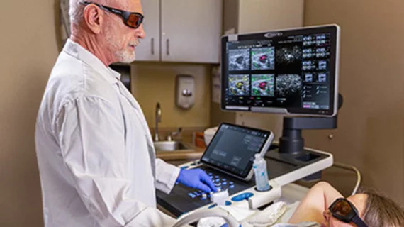
Emerging optoacoustic ultrasound technology promises to reduce false-positives and unnecessary biopsies
An up-and-coming ultrasound technology could help to significantly reduce the number of unnecessary biopsies of benign breast masses, according to a study published in the American Journal of Roentgenology.
For the study, researchers compared conventional ultrasound images to fused optoacoustic ultrasound images. Optoacoustic ultrasound (OA/US) fuses the anatomical features of a mass with functional data on what is occurring around the area of interest—neoangiogenesis, deoxygenation of hemoglobin, etc. OA/US can noninvasively provide valuable information on tumor behavior and better inform providers of whether a mass is benign or malignant.
Those additional details of tumor function are of value to clinicians because low specificity for differentiating between benign and malignant masses is associated with high rates of false positive reads on breast ultrasound exams and, in turn, increased rates of unnecessary biopsies, lead researcher Stephen Seiler, MD, with University Texas Southwestern Medical Center in Dallas, and colleagues explained.
“The potential harms of false-positive results on breast imaging examinations have garnered much attention, resulting in a drive to improve the specificity of imaging,” the experts noted. “This goal has led to increased utilization of functional imaging tools that provide supplementary biologic information to help differentiate benign masses from cancer.”
Researchers sought to determine whether the use of fused OA/US images would improve reader specificity at fixed sensitivity in comparison to conventional ultrasound images alone. To do this, the experts first had 15 readers assign a probability of malignancy (POM) to 480 breast masses classified as BI-RADS category 3 through 5 based on conventional ultrasound images and clinical history.
Following training in optoacoustic imaging interpretation, the readers evaluated fused ultrasound and optoacoustic images using Seno Medical’s Imagio Optoacoustic Ultrasound Breast Imaging System. Next they viewed POM scores derived from the imaging system's machine learning-based decision support tool before assigning their own final POM to each mass.
Using the OA/US images with DST assistance, readers recorded a specificity at fixed sensitivity of 98%—a significant improvement from conventional ultrasound images alone. Partial AUC was also higher across all readers for the fused OA/US images compared to conventional ultrasound.
Experts involved in the study suggested that the results of their study indicate a role for OA/US, integrated with DST assistance, in decreasing the number of biopsies owed to false positive interpretations derived from conventional breast ultrasound exams.
The study abstract is available in the American Journal of Roentgenology.

