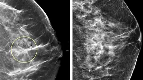
DBT not the superior modality for assessing BI-RADS 4 breast lesions
Though many institutions have migrated fully to digital breast tomosynthesis for routine breast cancer screening, new research suggests that in certain instances digital mammography remains the more effective imaging option.
In a modality comparison using more than 11,000 encounters of patients with BI-RADS 4-designated lesions, digital breast tomosynthesis exams showed no significant advantages over standard 2D mammography, researchers are reporting in a new study published in the European Journal of Radiology. While DBT exams were utilized more frequently than 2D studies included in the research, they did not yield significantly better results for breast cancer detection.
Experts sought to analyze the efficacy of both DBT and 2D studies specifically for the BI-RADS 4 category because of widely variable malignancy rates produced by the designation.
“Among the BI-RADS categories, BI-RADS 4—“suspicious findings with a recommendation for biopsy”—stands out for its enormous uncertainty, with a 2% to 95% likelihood of malignancy,” first co-author Chika F. Ezeana, of the Department of Systems Medicine and Bioengineering at Houston Methodist Cancer Center, and co-authors wrote. “A biopsy is thus considered as the most appropriate next course of action for BI-RADS 4 categorized lesions and serves as a quality metric and performance standard, resulting in a majority (69 to 95%) of BI-RADS 4 lesions being biopsied.”
The authors noted that biopsy-proven positive predictive values (PPV) for BI-RADS4 lesions have not improved throughout the years, despite the growing use of DBT, which has been shown to improve cancer detection in several studies.
For this reason, many breast specialists have begun to question whether DBT offers any significant advantages over 2D mammography, the authors suggested.
For the present study, researchers compared the findings from 6,356 2D and 5,896 DBT exams assessed as BI-RADS 4. After reviewing radiology reports and biopsy data, researchers found that while DBT studies were utilized 5.66% more often than 2D mammography, this did not increase cancer detection rates for the modality. In the DBT exams, the cancer detection rate was 120.76 compared to 112.65 for 2D. Positive predictive values were also similar, at 14.41% for 2D and 15.99% for DBT.
“Early studies have reported that DBT detects as much as 40% more cancers than digital (2D) mammograms in breast cancer screening examinations,” the authors stated. “Nevertheless, our findings indicate that once the assessment is made as BI-RADS 4, biopsy outcomes were comparable for both DM and DBT as malignancy rates, cancer detection rates, and biopsy-derived positive predictive values were similar, such that differences were not statistically significant.”
Although the results of the study did not highlight any significant advantage DBT use in BI-RADS 4, the authors noted that overall and in other BI-RADS categories, DBT is likely to perform better than standard 2D mammography.
Related breast imaging news:
Malignant architectural distortion ably diagnosed on breast imaging by human-AI combo
AI-based mammo screening protocol reduces radiologist workload by 62%

