Breast Imaging

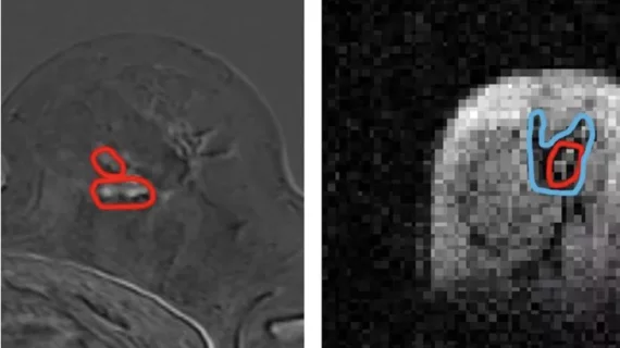
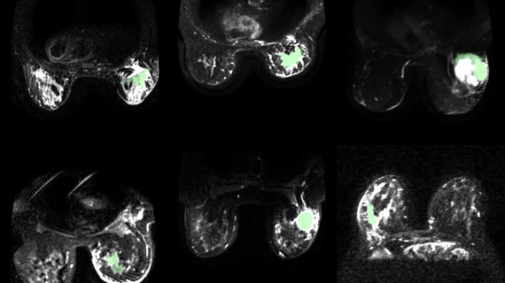
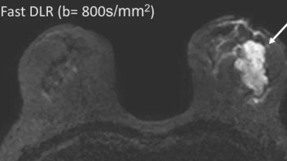
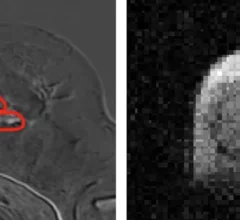
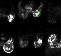
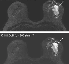
Breast imaging includes imaging modalities used for breast cancer screenings and planning therapy once cancer is detected. Mammography is the primary modality used. Mammogram technology is moving from 2D full-field digital mammography (FFDM) to breast tomosynthesis, or 3D mammography, which helps reduce false positive exams by allowing radiologists to look through the layers of tissue. Overlapping areas of dense breast tissue on 2D mammograms appear similar to cancers and 3D tomo helps determine if suspect areas are cancer or not. About 50% of women have dense breast tissue, which appears white on mammograms, the same as cancers, making diagnosis difficult. Radiologists use the Breast Imaging Reporting and Data System (BI-RADS) scoring system to define the density of breast tissue. Many states now require patients to be notified if they have dense breasts so they understand their mammograms might be suboptimal and they should use supplemental imaging that can see through the dense areas. This includes tomosythesis, breast ultrasound, automated breast ultrasound (ABUS), breast MRI, contrast enhanced mammography and nuclear imaging, including positron emission mammography (PEM).

Most popular radiology video interviews on Health Imaging. The evolving role of artificial intelligence (AI) in breast imaging was a big topic at the Radiological Society of North America (RSNA) 2024 meeting. Health Imaging spoke with Manisha Bahl, MD, breast imaging division quality director and breast imaging division co-service chief at Massachusetts General Hospital and an associate professor of radiology, Harvard Medical School, at the conference to hear more about her breast AI sessions.

Most popular radiology video interviews on Health Imaging

Radiology has seen a lot of large language model and generative AI research and adoption and it is clearly a hot button topic with our No. 1 video.

Manisha Bahl, MD, breast imaging division quality director and breast imaging division co-service chief, Massachusetts General Hospital, and an associate professor of radiology, Harvard Medical School, explains the findings of a recent study she was involved in at RSNA 2024. She also offers insights into growing interest at sessions in using AI in breast imaging.

What radiologists think about using ChatGPT and AI in breast imaging

Manisha Bahl, MD, explained that ChatGPT and other large language models offer significant potential to help radiologists with breast imaging exams, but they are "not quite ready for primetime."

FCI scanner more ably detects cancer spread than traditional MRI

New low-field scanner detects cancer spread better than traditional breast MRI

Researchers involved in its development are hopeful that the scanner could eventually lead to improved outcomes in cancer patients who require surgery to remove malignant tissue. 

CDI technique makes breast cancer glow on imaging.

MRI technique makes breast cancer 'glow' on imaging

The technique highlights differences in how water molecules move through cancerous tissue in comparison to healthy tissue.

Deep learning reconstruction improves breast MRI quality

Deep learning reconstruction cuts breast MRI scan times in half

The use of DLR also provides greater flexibility with protocols in comparison to conventional single-shot echo-planar imaging.

Breast cancer AI ribbon pink artificial intelligence

AI effectively flags mammograms of women who would benefit from supplemental MRI

Experts involved in the algorithm's development believe its time-saving potential could help improve both radiologist workflows and patient outcomes. 

Jessica H. Porembka, MD, FSBI, associate professor, breast imaging division University of Texas Southwestern Medical Center, Dallas, and vice chair of strategy and quality, and quality assurance medical director, Parkland Radiology in Dallas, explains how an ultrasound-first strategy for noncalcified lesions in DBT proves cost-effective.

Ultrasound-first strategy for noncalcified lesions in DBT proves cost-effective

Jessica Porembka, MD, of the breast imaging division at University of Texas Southwestern Medical Center, said an ultrasound-first strategy for these lesions in DBT is cost-effective and improves efficiency. 

Jessica H. Porembka, MD, FSBI, associate professor, breast imaging division University of Texas Southwestern Medical Center, Dallas, and vice chair of strategy and quality, and quality assurance medical director, Parkland Radiology in Dallas, discusses what it means when a radiology report says a patient has an architectural distortion on their mammogram.

What patients need to know about architectural distortion on breast imaging

Radiologist Jessica Porembka, MD, FSBI, an associate professor with the breast imaging division at University of Texas Southwestern Medical Center, explains what it means when a mammography report says a patient has architectural distortion.
 

Around the web

GE HealthCare designed the new-look Revolution Vibe CT scanner to help hospitals and health systems embrace CCTA and improve overall efficiency.

Clinicians have been using HeartSee to diagnose and treat coronary artery disease since the technology first debuted back in 2018. These latest updates, set to roll out to existing users, are designed to improve diagnostic performance and user access.

The cardiac technologies clinicians use for CVD evaluations have changed significantly in recent years, according to a new analysis of CMS data. While some modalities are on the rise, others are being utilized much less than ever before.