Microwave breast imaging: A non-invasive, non-ionizing emerging technology for breast cancer screening
Microwave breast cancer imaging is an emerging technology that offers patients in need of screening for breast cancer a non-invasive, non-ionizing option [1].
Microwave breast cancer imaging (MWI) devices make use of electromagnetic waves to penetrate breast tissue, which highlight differences in dielectric properties of healthy and cancerous tissue to identify abnormalities. Higher dielectric permittivity is more common in cancerous tissue in comparison to healthy glandular tissue.
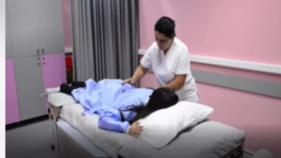
While MWI offers patients a non-invasive option for breast cancer screening (patients simply lay face down on an exam table with breasts inserted into the device through an opening in the bed), perhaps its most beneficial feature its ability to image the breast without the use of ionizing radiation, according to experts involved in a new study that tested the device’s utility in a screening capacity.
“This can have a major impact on the younger population, especially for women at high risk of developing breast cancer, like those with genetic mutations, where there is a necessity to start screenings at an early age,” corresponding author Ibrahim Akduman of Istanbul Technical University in Turkey and colleagues explained.
Akduman and colleagues recently tested the utility of their MWI-based SAFE (Scan and Find Early) device in conjunction with a machine learning approach (Adaptive Boosting model) on a cohort of 113 women with BI-RADS category 4 or 5. The patients were imaged with the device prior to undergoing biopsy. Biopsy results were used to compare the sensitivity, specificity and accuracy of the diagnoses derived from imaging.
For differentiating between benign and malignant lesions, the classification model achieved a sensitivity, specificity and accuracy of 79%, 77% and 78%, respectively. Lesions as small as 4 mm were ably detected using the device as well, suggesting utility for identifying cancers in earlier stages.
The MWI-based SAFE device was found to be more effective in younger patients. Outside of age, breast density also impacted detection rates, with higher density yielding lower detection of malignant lesions.
The researchers noted that the device is still under development, but that multicenter trials are in the works to test its clinical utility as a screening tool.
Learn more in Academic Radiology.

