Medical Imaging

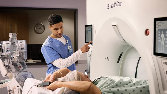
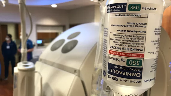
Physicians utilize medical imaging to see inside the body to diagnose and treat patients. This includes computed tomography (CT), magnetic resonance imaging (MRI), X-ray, ultrasound, fluoroscopy, angiography,  and the nuclear imaging modalities of PET and SPECT. 

Thumbnail

Differences in brain volume, gray matter thickness tied to epilepsy

New research from the University College of London (UCL) and the Keck School of Medicine at the University of Southern California (USC) found that epilepsy correlates with thickness and volume differences in the gray matter of several areas of the brain.

Images of newborns' brains could be key to detecting neurological disorders

In a few years, brain images of more than 1,000 newborns and another 500 fetuses will be generated through a high-resolution MR imaging project in hopes to find the source of neurological disorders at or before the time of birth, according to a recent article by the Financial Times.  

The new age of ultrasound

Ultrasound may not be as cutting-edge as, say, nuclear imaging or pack the punch of CT, but the technology that’s now more than a century old is making a comeback in unexpected avenues.

Thumbnail

Cause of alcohol dependence may be visible with molecular imaging

Researchers may have found a clearer understanding of what causes alcohol dependence, according to a new study published in the Journal of Nuclear Medicine (JNM).   

8 things to know about breast cancer screening for high-risk women

According to a recent study in the Journal of the American College of Radiology, the ACR recommends women at an average risk for breast cancer should begin annual mammograms at age 40. High-risk women should start mammograms even earlier. 

Cerveau Technologies signs research agreement with University of Pittsburgh for investigational PET agent

Cerveau Technologies Inc. today announced an agreement with the University of Pittsburgh to support multiple projects over the next several years. These research projects are for studies of an early stage imaging agent ([18F]MK-6240) to be used in Positron Emission Tomography (PET) scans for assessing the status and progression of neurofibrillary tangles (NFTs) in the brain. NFTs made up of aggregated tau protein are a hallmark of several neurodegenerative diseases, including Alzheimer’s disease.

Non-invasive blood test brings physicians one step closer to detecting cancer early

Researchers may be closer to detecting cancers before symptoms become apparent in patients, according to a recent article by NPR. Although more research needs to be conducted, the newly developed non-invasive blood test would detect eight common types of cancer, with the potential to open up possibilities in the field of oncology.  

Thumbnail

Breast cancer survivors reveal the best way to follow up with them

A recent study from the American Journal of Roentgenology (AJR) has found imaging surveillance follow-up appointments vary among breast cancer patients and survivors nationwide, suggesting a strong need for refinery and consistency.

Around the web

RBMA President Peter Moffatt discusses declining reimbursement rates, recruiting challenges and the role of artificial intelligence in transforming the industry.

Deepak Bhatt, MD, director of the Mount Sinai Fuster Heart Hospital and principal investigator of the TRANSFORM trial, explains an emerging technique for cardiac screening: combining coronary CT angiography with artificial intelligence for plaque analysis to create an approach similar to mammography.

A total of 16 cardiology practices from 12 states settled with the DOJ to resolve allegations they overbilled Medicare for imaging agents used to diagnose cardiovascular disease.