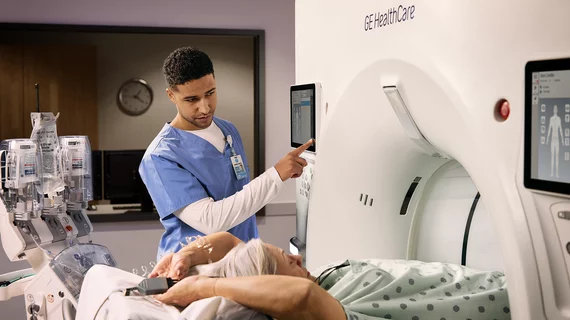
Medical Imaging

Physicians utilize medical imaging to see inside the body to diagnose and treat patients. This includes computed tomography (CT), magnetic resonance imaging (MRI), X-ray, ultrasound, fluoroscopy, angiography,  and the nuclear imaging modalities of PET and SPECT. 

Cancer overdiagnosis: A path forward

As oncologists, radiologists and epidemiologists continue to debate the details of overdiagnosis and overtreatment, a viewpoint published online July 29 in JAMA listed several recommendations for the National Cancer Institute to consider as it attempts to evaluate the overdiagnosis issue.

F-18 FDM may offer new oncologic PET agent

A fluorine-based PET agent on the workbench called 2-Deoxy-2-18F-fluoro-D-mannose (F-18 FDM) has been found to work similarly and potentially better than FDG for cancer imaging, especially of the brain, according to a study published July 10 in the Journal of Nuclear Medicine.

FDG PET/CT reveals necrotic nuances in sarcomas that predict survival

Little improvement in survival has been made for patients with sarcomas, despite an array of aggressive treatments. Researchers are trying to identify patients at high risk of recurrence by performing FDG PET/CT to quantify necrotic areas of tissue, a strong indicator of survival, according to research published in the July issue of the American Journal of Roentgenology.

USPSTF gives nod to CT lung cancer screening

The U.S. Preventive Services Task Force (USPSTF) has issued a Grade B recommendation for annual low-dose CT screening for individuals between the ages of 50 and 80 who are at high risk for lung cancer.

ED x-rays for kids with asthma spike

Emergency department (ED) use of chest x-rays for children presenting with moderate to severe asthma increased significantly from 1995 to 2009. Pediatric EDs bucked the trend, indicating a need to transfer best practices to standard EDs, which could improve efficiency, cut costs and decrease radiation exposure, according to a study published in the August issue of Pediatrics.

Mobile imaging center supports riders on cycling’s biggest stage

In the Tour de France, every minute matters. For riders who sustain what could be a serious injury, the only way to be evaluated and cleared to continue riding would be to travel off-site to a hospital.

PET flexes its muscles

The power of PET came across loud and clear in July. Studies showed its utility in informing management of triple-negative breast cancers, monitoring Alzheimer’s progression and predicting chemo success for patients with non-small cell lung cancer.

Thumbnail

MR + PET highlight elusive triple negative breast cancers

The heterogeneity of breast cancer can make it difficult to capture, especially in cases where telltale receptor expression or tumor characterization are not involved. However, MR and F-18 FDG PET may be able to suss out the more obscure phenotypes of the disease, according to a study published July 17 in Radiology.

Around the web

RBMA President Peter Moffatt discusses declining reimbursement rates, recruiting challenges and the role of artificial intelligence in transforming the industry.

Deepak Bhatt, MD, director of the Mount Sinai Fuster Heart Hospital and principal investigator of the TRANSFORM trial, explains an emerging technique for cardiac screening: combining coronary CT angiography with artificial intelligence for plaque analysis to create an approach similar to mammography.

A total of 16 cardiology practices from 12 states settled with the DOJ to resolve allegations they overbilled Medicare for imaging agents used to diagnose cardiovascular disease.