AI, imaging project to improve precision medicine for treating concussions
Researchers involved in a national multi-year, multi-center project are challenging the U.S. Department of Energy's supercomputers while analyzing brain imaging data via artificial intelligence (AI) to improve treatment of traumatic brain injury (TBI), according to a July 3 press release.
Since March, researchers from national laboratories and various academic institutions around the country have analyzed TBI data from 3,000 patients, including advanced CT and MRI, proteomic and genomic biomarkers, and clinical outcomes using AI-based technologies and the nation’s top supercomputing resources, according to the release.
“By harnessing subject matter expertise across the participating universities and labs and using next-generation AI-based tool development for precision diagnostics, we are optimistic that we will be able to create clinically actionable information to guide personalized treatment for TBI patients,” said study researcher Geoff Manley, MD, PHD, contact principal investigator and professor of neurosurgery at the University of California, San Fransisco (UCSF), in a prepared statement.
The study aims to bring precision diagnostics and prognostic modeling to TBI via a large dataset for application in clinical settings, explained study researcher Shankar Sundaram, MD, an engineer at Lawrence Livermore National Laboratory.
“We’ll need to be able to build suitable data infrastructure to transfer, store and provide compute access to this complex data at high speeds and efficiencies, and develop new algorithms and workflows to process multimodal data, such as images, genomic and biomarker information, together with detailed outcome assessments and learn patterns," Sundaram said. "An additional challenge is to understand how to arrive at diagnostics in the context of missing and uncertain data, and it has to be interpretable, so we can explain (to the clinician) what the model is telling them and why.”
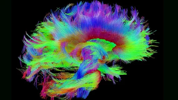
Using Department of Energy supercomputers, researchers were able to create a “connectome,” or a model of the structural connectivity of the human brain from MRI data in a matter of hours.
Also, by utilizing data from the 3,000 TBI patients gathered from a consortium led by UCSF, researchers will attempt to create a streamlined system for sharing massive amounts of research data among the Department of Energy laboratories, according to the release.
They will then develop a predictive model that could be used to triage patients into risk categories and assess potential outcomes, according to the release and, if successful, could inform future models of other diseases or disorders.
"This type of project is possible only when we bring together the new levels of computing capability available in Department of Energy with the revolutionary new analytics being developed through machine learning,” said Jim Brase, the principal investigator on the project. “It takes both elements to make progress. This is one of the most difficult computing challenges we’ve ever seen.”

