Radiologists use diagnostic imaging to non-invasively look inside the body to help determine the causes of an injury or an illness, and confirm a diagnosis. Providers use many imaging modalities to do so, including CT, MRI, X-ray, Ultrasound, PET and more.
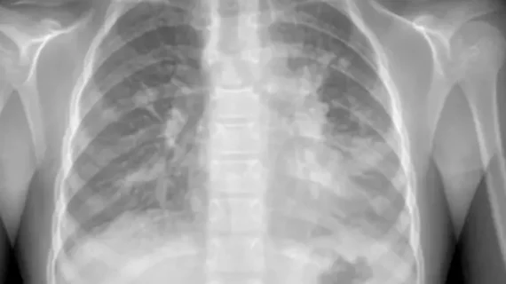
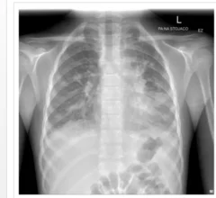
Following a recent surge of mycoplasma pneumoniae pneumonia cases, experts have issued new guidance to help providers quickly identify and treat the condition, with imaging playing a prominent role.
Following a recent surge of mycoplasma pneumoniae pneumonia cases, experts have issued new guidance to help providers quickly identify and treat the condition, with imaging playing a prominent role.
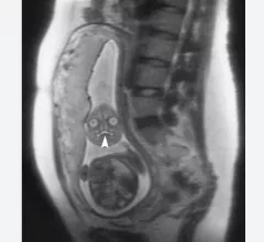
This is a clinical photo gallery of fetal imaging that explains what all can be seen on medical imaging, how sex is determined, how measurements are used to track the development of a baby.
Serious discrepancies between preliminary imaging reads and final radiology reports are at risk of accumulating when the prelims are rendered during overnight hours.
Clinicians have been using HeartSee to diagnose and treat coronary artery disease since the technology first debuted back in 2018. These latest updates, set to roll out to existing users, are designed to improve diagnostic performance and user access.
The cardiac technologies clinicians use for CVD evaluations have changed significantly in recent years, according to a new analysis of CMS data. While some modalities are on the rise, others are being utilized much less than ever before.
The new guidelines were designed to ensure sonographers and other members of the heart team have the information they need to screen patients when appropriate and identify early warnings signs of PH.