Researchers advocate for stricter chest x-ray imaging for children with epicardial pacemakers
Researchers from Boston Children’s Hospital and Harvard Medical School recommend stricter chest x-ray screenings of children fitted with epicardial pacemakers to detect those at risk of coronary artery compression, according to a study published online Aug. 13 in the journal HeartRhythm.
"Coronary artery compression is thought to be rare," lead author Douglas Mah, MD, director of the Pacemaker and ICD Program at Boston Children's Hospital and assistant professor of pediatrics at Harvard Medical School, said in a prepared statement. "Its true incidence, however, may be higher than we believed due either to a lack of awareness or lack of reporting in the literature."
Mah and colleagues reviewed 145 patients' records archived at Boston Children’s Hospital from 2000 through 2017 who had either active or epicardial wires that included coronary imaging, either CT or angiography. The researchers found 5.5 percent of patients showed some level of coronary compression from their epicardial pacemakers, which resulted in seven patients having undergo surgery for device removal or repositioning.
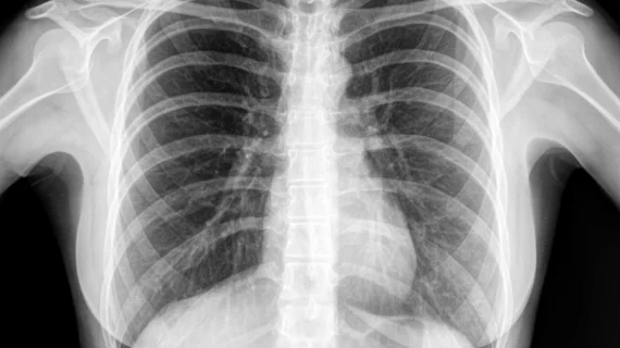
As position of epicardial device wires may change as the child grows, Mah and colleagues recommended that chest x-rays should be conducted every few years.
However, if the child has symptoms such as unexplained chest pain, fatigue, or evidence of heart muscle damage or dysfunction, a cine CT scan should be used due to the its low radiation and non-invasive properties. A last resort is to have the patients undergo catheter angiography to confirm diagnosis before surgery, according to the researchers.
"The use of pacemakers and defibrillators in children is growing—as more epicardial devices are implanted, more children may be at risk for developing coronary compression from their leads,” Mah said. “We hope to increase awareness among healthcare providers and patients of this important, possibly preventable, and potentially fatal complication and provide a useful screening algorithm to detect at-risk patients and ultimately prevent complications."

