CT technique offers microscopic view of Egyptian remains
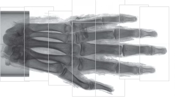
Swedish researchers have used a novel CT imaging technique to study the soft tissue of an ancient Egyptian mummy’s hand at microscopic levels, according to a Sept. 25 study published in Radiology.
X-ray and CT imaging have both advanced archaeology and paleopathology, allowing scientists to analyze diseases embedded within mummified soft tissue. However, the conventional modalities provide low observable resolution, forcing researchers to use invasive and often destructive methods.
"For studying bone and other hard, dense materials, absorption contrast works well, but for soft tissues the absorption contrast is too low to provide detailed information," said Jenny Romell, from KTH Royal Institute of Technology/Albanova University Center in Stockholm, Sweden, in a news release. "This is why we instead propose propagation-based phase-contrast imaging."
The proposed method works by detecting absorption and phase shift that happens when x-rays move through a sample—thus providing higher contrast for soft tissues.
Romell and colleagues used laboratory phase-contrast CT arrangement with propagation-based imaging on a mummified human right hand from ancient Egypt. They imaged the hand using two different modes: a CT scan of the whole hand and a detailed scan of the tip of the middle finger.
With an estimated resolution between six to nine micrometers—just wider than a human red blood cell—the team visualized remains of adipose cells, blood vessels and nerves. Romell et al. also observed blood vessels in the nail bed and various layers of skin in the hand.
"With phase-contrast CT, ancient soft tissues can be imaged in a way that we have never seen before," Romell added.
As a result of their findings, the group believes phase-contrast CT may be a key addition or replacement to the soft-tissue extraction and chemical processing methods currently used by paleopathologists, they wrote.
"Just as conventional CT has become a standard procedure in the investigation of mummies and other ancient remains, we see phase-contrast CT as a natural complement to the existing methods," Romell said. "We hope that phase-contrast CT will find its way to the medical researchers and archaeologists who have long struggled to retrieve information from soft tissues, and that a widespread use of the phase-contrast method will lead to new discoveries in the field of paleopathology."

