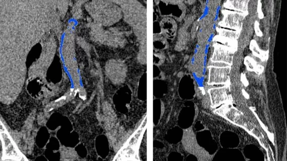
CT reveals abdominal aortic calcification can predict future heart attack, stroke
Heart attack and other life-threatening cardiovascular events can be predicted with CT-based measures of calcification in the abdominal aorta and could be a more accurate predictive model than the widely known Framingham risk score, according to research published Oct. 2 in Radiology.
"We found a strong association between abdominal aortic calcification and future cardiovascular events," said lead author Stacy D. O'Connor, MD, assistant professor of radiology at the Medical College of Wisconsin, in a prepared statement. "With heart disease being a leading cause of death, anything we can do to make our patients more aware of their risk is going to help."
Although the Framingham risk score can help predict cardiovascular disease based on traditional risk factors (age, cholesterol, blood pressure, etc.), many patients who fall into an indeterminate risk category could benefit from additional noninvasive assessment methods such as CT imaging to measure calcium levels in the coronary arteries.
For their study, O’Connor and colleagues evaluated the relationship between abdominal aortic calcification on CT and cardiovascular events in 829 asymptomatic patients at an average age of 58 years. The study cohort had undergone CT colonography between April 2004 and March 2005.
The researchers then assessed the patients for an average time period of 11 years to determine if they developed adverse cardiovascular events such as heart attack, stroke, death or congestive heart failure—and of the 829 patients, 18.8 percent did.
The researchers noted that major cardiovascular events occurred an average of seven years after CT and included heart attack in 39 patients and death in 79. Overall, CT based abdominal aortic calcification was five times higher in those who had a cardiovascular event than those who didn’t, and ultimately outperformed the Framingham risk score, according to the researchers.
CT abdominal aortic calcification assessment could be a valid screening tool added to other cardiovascular risk exams without the need for additional patient time and radiation dose, the researchers added.
"There are thousands of CT scans performed every day across the United States, so this gives us an opportunity to reach a lot of people," O'Connor said in the same statement. "For instance, if someone is getting a scan for cholecystitis and we see abdominal aortic calcification on the CT, we can address things like blood pressure and cholesterol with the patient."

