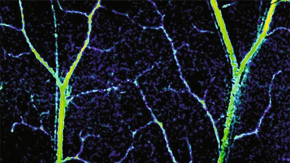
Novel 3D imaging tool captures blood flow in the capillaries
Researchers at Northwestern University in Evanston, Illinois have developed a noninvasive, three-dimensional (3D) imaging tool able to capture blood flow and oxygenation within the capillaries of a human, according to research published online Jan. 16 in the journal Light: Science & Applications.
Capillaries are responsible for delivering oxygen to organs and tissues through the body while ridding it of unnecessary carbon dioxide. However, low blood oxygen can cause a variety of conditions, from headaches to cardiovascular disease.
Imaging techniques such as doppler ultrasound, which is used to see inside major veins and arteries, can’t fully image the circulatory system due to the microscopic size of capillaries. Instead, the researchers’ technique can detect alterations to capillary organization and may lead to earlier diagnosis and treatment.
“There has been a progressive push to image smaller and smaller blood vessels and provide more comprehensive, functional information,” lead author Vadim Backman, PhD, a professor of biomedical engineering at Northwestern’s McCormick School of Engineering, said in a prepared statement. “Now we can see even the smallest capillaries and measure blood flow, oxygenation and metabolic rate.”
Called spectral contrast optical coherence tomography angiography (SC-OCTA), the tool is a 3D imaging technique which can detect tiny changes in capillary organization and indicate early signs of diseases to prompt earlier diagnosis.
SC-OCTA combines spectroscopy—which looks at various visible light wavelengths—with conventional optical coherence tomography (OCT). In short, OCT identifies the tissue of interest and then spectroscopy characterizes it.
The technique also does not require dyes, radiation and can image stagnant blood or moving organs—regardless of its speed—such as a beating heart.
“SC-OCTA’s unique ability to image non-flowing blood could also become a valuable tool for the booming field of organoids, which studies how organs develop and respond to disease,” said first author James Winkelmann, a biomedical engineering graduate student at Northwestern.
The technology does have its limitations as it currently can’t image deeper than one millimeter, which is less than the depth an ultrasound can image. The researchers are working to put the tool on the end of an endoscopic probe so it can be inserted into the body and image organs up-close.
The study was supported by the National Science Foundation and the National Institutes of Health.

