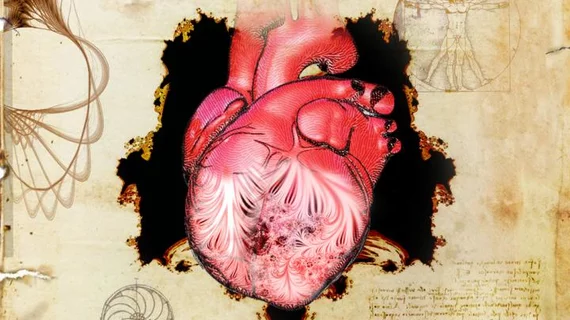
Thousands of MRI scans help experts understand 500-year-old heart mystery first described by Leonardo da Vinci
In the 16th century, Leonardo da Vinci first sketched intricate patterns within the heart that he believed were important to its functioning. And now, 500 years later, a team of experts has turned to medical imaging for an answer.
Collaborators from a number of international centers, led by the European Molecular Biology Laboratory-European Bioinformatics Institute, published their findings Wednesday in Nature. Using MRI scans, along with morphology and genetic data, they showed that this network of muscle fibers—known as the trabeculae—can influence the heart’s performance.
Artificial intelligence and more than 25,000 cardiac MRIs also helped the group determine that the development of these muscles affects an individual’s risk of heart failure.
"Our findings answer very old questions in basic human biology,” Deputy Director General of EMBL, Ewan Birney, said in a statement. “As large-scale genetic analyses and artificial intelligence progress, we're rebooting our understanding of physiology to an unprecedented scale.”
Trabeculae begin to take shape early in human development, with the geometric patterns providing oxygen as the heart grows, the authors wrote. Centuries ago, da Vinci thought this network warmed the blood as it moved through the vital organ, but it’s long remained a mystery.
Birney and colleagues determined that it’s the shape of the trabeculae that influences cardiac function, which they also believe points to a potential link to heart disease. The group confirmed their hypothesis by analyzing genetic information from some 50,000 patients.
They also pinpointed six regions in human DNA that can impact how these fractal patterns form.
“Leonardo da Vinci sketched these intricate muscles inside the heart 500 years ago, and it's only now that we're beginning to understand how important they are to human health,” Declan O'Regan, clinical scientist and consultant radiologist at the MRC London Institute of Medical Sciences, said in a statement.
The MRC London Institute of Medical Sciences, Heidelberg University, and the Politecnico di Milano all contributed to this research.

