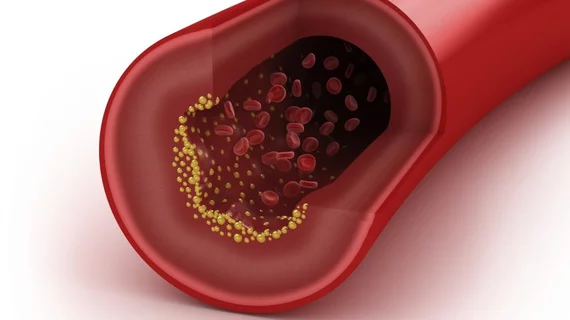
CT reveals HIV patients living with higher levels of dangerous coronary plaque
CT scans of patients with HIV and without known cardiovascular disease revealed higher levels of dangerous coronary plaque compared to those without the virus, according to new research published in Radiology.
In fact, exams revealed HIV patients have 2-3 times the noncalcified plaque burden as healthy volunteers, physicians reported Tuesday. Such buildup is more prone to rupture compared to calcified plaque and underscores the need for these patients to live a heart-healthy lifestyle.
Study author Chartrand-Lefebvre, MD, MSc, and colleagues suggest those with HIV understand their cardiovascular risk factors such as smoking, diabetes, high blood pressure, obesity and a sedentary lifestyle. The findings also have important implications for imaging providers, they noted.
“For radiologists, these results suggest that coronary CT angiography interpretation in people living with HIV should probably include quantification of coronary plaque by subtypes to allow better cardiovascular risk stratification,” Chartrand-Lefebvre, with the Radiology Department at the Centre hospitalier de l'Université de Montréal in Montreal, added in a statement.
To arrive at their conclusions, the authors prospectively compared coronary plaque burden and CT characteristics of 265 participants, including 181 asymptomatic individuals with HIV and without CAD, along with 84 completely healthy volunteers.
Radiologists blinded to participants’ clinical history and HIV status used CT angiography results to assess plaque. Noncalcified prevalence and volume were higher in those with HIV, even after adjusting for cardiovascular risk factors.
This is likely due to many variables, the authors noted, including the use of antiretroviral therapy, which does not cure the disease but helps control it and has significantly contributed to lowering HIV mortality rates.
Of note, there was no difference between the two cohorts in terms of coronary artery calcium score, overall plaque prevalence, and 10-year Framingham Risk Score. The latter is often used to predict the risk of CAD, the authors explained.
Read the entire study here.

