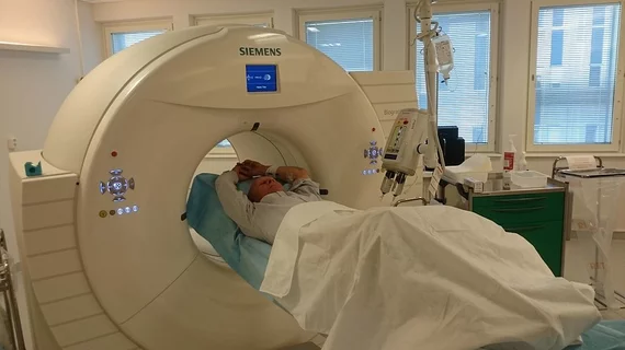
Simple, proven strategies to reduce extravasation of contrast media during CT scans
The risk of contrast media extravasation during CT scans can be reduced when venous assessments and prevention protocols are implemented in radiology departments.
A retrospective analysis of more than 73,000 patients who underwent contrast-enhanced CT scans revealed that interventions to prevent such IV leakages produced measurable reductions in incidents.
The practical solutions to reduce the instances of extravasation of contrast media (ECM) were published recently in Academic Radiology. There, researchers shared how to best prevent the complication when administering contrast media intravenously, as well as why it is important for the radiology team to be vigilant in doing so.
“Subcutaneous extravasation of contrast media is a well-known complication during contrast-enhanced computed tomography scans, occurring even when appropriate intravenous injection techniques are used,” corresponding author Seitaro Oda, MD, with the department of diagnostic radiology at Kumamoto University, and co-authors explained. “The majority of ECMs reportedly involve small volumes of contrast media and resolve without severe adverse effects. However, severe complications, such as tissue necrosis, neurovascular compromise, or compartment syndrome, can potentially occur.”
At the researchers’ institution, strategic venous assessments before IV contrast administration began in January 2015. The assessments were performed by radiology nurses, who would examine patients’ veins for fragility, swelling, age and the presence or absence of blood regurgitation into the IV access tube. If an issue was encountered, the nurses consulted with radiologists, who then decided how to proceed based on that patient’s situation.
Their prevention scan protocols were introduced in January 2017. Protocols were based on the venous assessments and included decreasing contrast media injection rates and having the CT technologists complete the scans using low tube voltage imaging. The contrast was warmed in all cases and, when necessary, nurses were on standby to monitor the IV site.
When analyzing thousands of records for the CT scans that were completed between January 2013 and December 2019, researchers examined how the protocols impacted extravasation rates before, during and after interventions.
Out of the 73,931 patients, ECM occurred in .39% (292) and the researchers found that their interventions played a significant role in reducing extravasation. For periods A (no prevention), B (venous assessment only) and C (venous assessment and scan protocol implementation), the ECM rates were 0.62%, 0.43% and 0.24%, respectively.
“Preventive strategies were effective for reducing the frequency of ECM during CT,” the doctors wrote. “The strategies are considered to be simple and clinically practical, and the radiology team (radiology nurses, radiology technicians, radiologists) can play an important role in preventing ECM during CT.”
More on contrast media:
Allergic reactions to iodinated CT contrast increase likelihood of sensitivity to GBCAs
Warming up CT contrast agents raises radiology department costs and few clinical benefits
Guerbet, Bracco partner on new lower dose gadolinium contrast agent for MRI scans
Fewer than 2% of patients know which imaging contrast caused their allergic reaction

