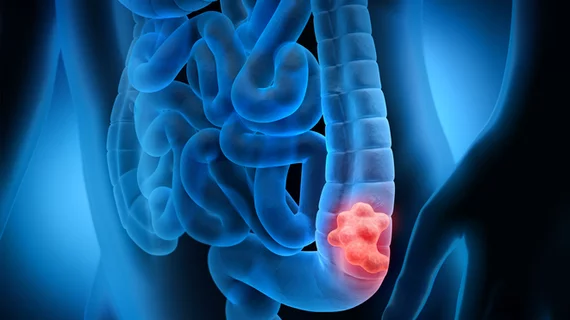
CT-based radiomics nomogram accurately predicts colorectal cancer prognosis
Researchers developed a nomogram that uses a CT-based radiomics signature to accurately predict overall survival in colorectal cancer patients.
When combined with clinical predictors and genetic characteristics (KRAS/BRAF mutation status), the signature improved the predictive accuracy for postoperative prognosis in CRC patients, and researchers involved in the study suggested that their findings could impact treatment strategies.
“The results showed that CT-based radiomics features not only have potential predictive value for overall survival in CRC patients, but also that the combined models of CT-based radiomics features and pathological T staging and KRAS/BRAF gene phenotypes can further improve the predictive power of overall survival in CRC patients,” corresponding author Feng Feng, with the Department of Radiology at Affiliated Tumor Hospital of Nantong University in Nantong, China, and co-authors discussed.
Colorectal cancer incidence and mortality rates have increased significantly over the last several years. With the five-year-survival rate at around 14%, the need for an accurate prognosis-predictive model that could help determine mortality risk is urgent, as it could serve as a guiding tool for treatment decisions. Enhanced CT is the standard for diagnosing, staging and monitoring CRC. Radiomics can take imaging a step further by extracting high dimensional features of tumors to quantify the information and use it to improve the clinical prognosis. When combined, radiomics and CT imaging can further boost prediction accuracy.
Researchers used imaging from 121 CRC patients to create their model, in addition to 51 and 114 patient exams selected for internal and external validation. From these images, radiomics features were extracted using various methods.
Researchers found that the radiomics features had a significant correlation with CRC patients’ survival time. The nomogram’s c-indexes for the training, internal validation and external test cohorts were 0.782, 0.721 and 0.677. When clinical predictors were integrated with the signature, the prediction accuracy improved further, with calibration curves revealing predicted survival times were very close to actual survival times.
“This demonstrates the reliability and reproducibility of the developed comprehensive nomogram model, and the potential value of radiomic signatures in assessing patient prognosis, enabling better generalizability of the findings of this study,” the authors wrote. “These findings might affect treatment strategies and enable a step forward for precise medicine.”
The detailed research can be viewed in Academic Radiology.
More on oncology imaging:
These ultrasound features predict ovarian cancer
Is MRI a suitable alternative to CT for testicular cancer surveillance? Research offers insight
Multimodal AI platform can accurately diagnose and stage thyroid cancer via ultrasound images
Breast MRI screening cuts cancer mortality rates in half for women with lesser-known gene mutations

