MRI a useful tool for avoiding invasive procedures for suspected idiopathic intracranial hypertension
Combining MRI findings with neurological and ophthalmological data can lead to more accurate diagnosis of idiopathic intracranial hypertension.
Overdiagnosis of idiopathic intracranial hypertension (IIH) has been reported in prior research to be as high as 39% in patients assessed for the disease. This could potentially be due to the overutilization of tests and procedures that are not necessary, corresponding author of a new European Journal of Radiology paper Beyza Nur Kuzan, from the Department of Radiology at the Kartal Dr. Lütfi Kırdar City Hospital in Istanbul, and colleagues recently explained [1].
“With the widespread use of cranial MRI, the incidence of incidentally detecting findings that may be related to IIH has increased,” the authors wrote, “which leads to further investigation of cases for IIH.”
What they did:
The researchers involved in the study sought to uncover whether specific imaging features, when combined with the neurological and ophthalmological data, of patients with suspected IIH could be indicative of disease. To do this, they analyzed the data from 98 patients—49 with IIH and 49 controls with similar demographic characteristics—who presented to their hospital with symptoms between January 1, 2018 and March 15, 2020.
What they found:
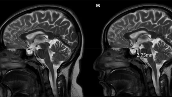
In terms of imaging features, lateral ventricular index yielded the highest AUC (.94) for prediction of disease, followed by sella height category (0.91) and optic nerve tortuosity (0.85). Using the multivariate model developed by the researchers, they also found that lower caudate index, lateral ventricle index and bilateral optic nerve tortuosity to be significant predictors of disease.
The experts maintained that these MRI parameters support the diagnosis of IIH in clinically suspicious cases and could help reduce the use of invasive procedures, such as lumbar punctures, when aligned with IIH symptoms.
The authors wrote:
A holistic approach to the clinical and radiological findings of the cases in the diagnosis of IIH may prevent overdiagnosis by providing the opportunity for early correct diagnosis. Future further studies implementing objective neuroimaging criteria, such as these quantitative measures, may hopefully aid in avoiding invasive procedures, such as recurrent LP, in the diagnostic evaluation of these patients.”
The study details can be viewed here.

