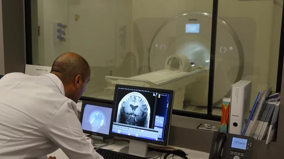
Time-of-flight intracranial MRA at 5T comparable to 7T, new analysis shows
Experts recently determined that three-dimensional time-of-flight MR angiography on a 5T system yielded results comparable to those produced by a 7T.
A new paper published in Radiology details an comparison of 3T, 5T and 7T to determine which system could achieve the best intracranial vascular image quality. The authors of the paper explained that although these images have been reported to be high quality at 7T, its application is limited.
“Three-dimensional (3D) time-of-flight (TOF) MR angiography (MRA) at 7T has been reported to have high image quality for visualizing small perforating vessels. However, B1 inhomogeneity and more physiologic considerations limit its applications. Angiography at 5-T may provide another choice for intracranial vascular imaging,” corresponding author of the paper Mengsu Zeng, from the Shanghai Institute of Medical Imaging in China, and co-authors explained.
For their research, the experts evaluated the imaging of 12 participants (some healthy, some with a history of ischemic stroke) who had undergone 3T, 5T and 7T 3D TOF MRA with use of customized MR protocols within 48 hours. Radiologists graded image quality and the Friedman test was used to compare the characteristics of each scan.
The distal arteries and small vessels at 5T TOF MRA were visualized significantly better than at 3T. Additionally, the total length of small vessel branches was reported to be larger at 5T than 3T. The experts did not note any significant differences between the images acquired at 5T compared to 7T.
These findings prompted the experts to suggest that angiography at 5T could offer a suitable alternative to using 7T systems.
The study abstract can be viewed here.

