Interactive teaching app improves reader performance on prostate MRI
An interactive educational app for medical students and radiology trainees can be utilized to improve users’ knowledge and understanding of prostate cancer on MRI exams.
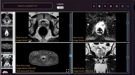
The LearnRadiology app was recently developed by a group of experts at the University of Chicago. When recently tested by one radiologist and two radiology residents, the teaching app was found to improve readers’ performance in detecting prostate cancer on multiparametric MRI (mpMRI) scans, encouraging its developers in regard to its future utility.
Details of the app and its resultant success among users were published May 1 in Academic Radiology.
Experts involved in the app’s creation suggested that because it was designed to mimic real life, its use among emerging radiologists could potentially help address the issue of subjectivity and reader variability in interpreting mpMRI prostate exams—something that other 3D interactive e-learning resources have not yet successfully addressed.
“The current modules that teach MRI still lack the application to clinical settings that include the mpMRI approach and the content necessary to improve the user’s visual memory and spatial ability for PCa diagnosis,” corresponding author of a new paper Aritrick Chatterjee, PhD, with the Department of Radiology at the University of Chicago, and colleagues explained. “Therefore, in this study, we aimed to create an interactive application with mpMRI—whole-mount histology correlation for enhanced prostate MRI training of radiologists.”
The app combines prostate MRI images with whole-mount histology for 20 cases curated for unique pathology and teaching points. To test the app, experts first had readers review 20 mpMRI prostate exams to identify areas of suspected cancer and provide a confidence score in their interpretation (without using the app). One month after using the app, users repeated the process on 20 different exams.
Upon comparing readers’ interpretations from before and after utilizing the app, the group noted a significant improvement in performance. Sensitivity and positive predictive value improved for all three readers after using the app, as did confidence scores for true positive cancer lesions.
“A critical part of the learning experience was to create a curated data set with teaching points to guide less experienced radiologists, mainly residents and fellows, to detect the abnormality or its absence after reviewing the learning app and integrating the information on these data sets to make the diagnosis,” the group noted. “LearnRadiology offers this experience by providing the entire data set of images that a trainee will encounter in practice.”
In the future, the team plans to explore additional areas that could improve users’ experience, including adding more curated cases with different pathologies, the use of AI and slides of different imaging modalities.
Learn more about the group’s work here.

