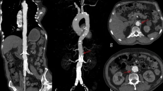
Deep learning reconstruction cuts radiation and contrast dose by half in aortic CTA exams
Deep learning image reconstruction (DLIR) techniques allow techs to reduce both contrast dosage and radiation exposure during aortic computed tomography angiography (CTA) exams, new data show.
Researchers recently discovered that, with the help of a deep learning reconstruction algorithm, they could reduce tube voltage by nearly half without sacrificing image quality. What’s more, they achieved diagnostic quality exams with significantly lower contrast dosage.
The group detailed their research Nov. 13 in Academic Radiology, where they explained the importance of finding balance between reducing radiation exposure and maintaining image quality.
“A significant drawback of reducing the tube voltage is the increase in image noise due to insufficient photon flux,” corresponding author Jie Liu, with the Department of Radiology at The First Affiliated Hospital of Zhengzhou University in China, and colleagues explained. “To mitigate this issue, the hybrid iterative reconstruction algorithm and deep learning image reconstruction algorithm are commonly used, as both can effectively suppress quantum noise and improve the signal-to-noise ratio.”
DLIR is known to help with reducing image noise while preserving high contrast spatial resolution and has been successfully deployed alongside lower kVp. The team sought to determine whether tube voltage as low as 60 kVp could be achieved during aortic CTA while also reducing contrast dosage.
To do this, researchers conducted a phantom analysis to develop imaging protocols using three different reconstruction techniques. A total of 90 participants set to undergo aortic CTA were recruited and then divided into three groups—one conventional group scanned using standard protocols, one imaged at 60 kVp with 105 mg iodine/kg with hybrid iterative reconstruction (60 kVp-CV group) and one using that same reduced protocol but with the addition of DLIR (60 kVp-CI group).
Overall, the team was able to reduce radiation and contrast dose by 45% and 50% in the experimental groups. In the experimental group that had DLIR applied to their imaging, the CT attenuation, SNR and CNR were superior or equal to that observed in the group that underwent the standard protocol. Image quality was comparable between both groups that underwent the reduced protocols and the DLIR group’s images were deemed sufficient across all vascular segments.
“These results indicate that the scanning protocol using 60 kVp and reduced CM dosage, combined with DLIR-CI algorithm, is effective for low-dose aortic CTA examinations,” the group noted. “This finding is clinically significant as it demonstrates the potential for reducing both radiation and CM dosages while maintaining diagnostic image quality.”
One possible limitation to the effectiveness of DLIR is that it may not be as effective in patients who are obese, particularly within the abdominal region, the authors explained. To compensate, the team suggested that the algorithm could be successfully implemented with certain restrictions based on abdominal circumference.
The study abstract is available here.

