What’s more, use of the AI software also resulted in improved sensitivity and specificity, according to new research published in Insights into Imaging.
CT value difference between pre-surgery images and uncorrected post-surgery images, post-surgery O-MAR images, and post-surgery dl-MAR measured in ROIs on six different locations. Credit: European Journal of Radiology
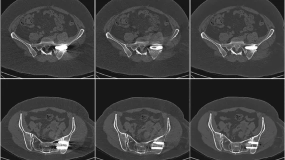
This is the first study to use paired, real-world clinical CT images to evaluate deep learning-based artifact reduction techniques.
Rather than administering radiolabeled glucose for exams, imagers give patients a small amount of a harmless glucose solution that is said to be equivalent to a can of a carbonated drink.
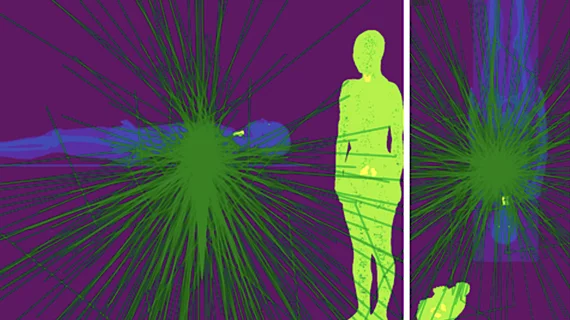
X-ray photon trajectory during the simulation phantom study from the side and top views. Due to scattering of the X-rays when they hit the lower end of the patient bed, exposure in mainly to the lower body of the interventional echocardiographer performing transesophageal echocardiography. The green lines are the scattered photon trajectories calculated by Monte Carlo simulation in the study.
Researchers found that echocardiographers in the cath lab are exposed to high doses of radiation on the right half of their body, especially the waist and lower body.
Difficulty obtaining PET/CT scans was reported by 55% of respondents, with 21% citing this as the greatest barrier in treating classic Hodgkin lymphoma.
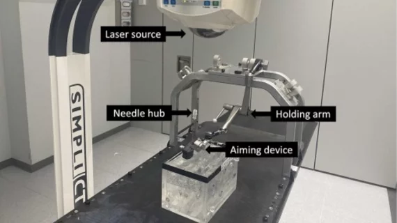
Setup showing the laser guidance system in combination with the aiming device. Credit: Academic Radiology.
Laser guidance has been shown to improve needle placement accuracy during percutaneous interventions, but this procedure has drawbacks, experts explained recently.
Example of interventional echocardiography TEE imaging superimposed on live fluoro during a transseptal puncture for a MitraClip procedure.
The American Society of Echocardiography (ASE) released a new guideline document that outlines uniform training standards for interventional sonographers guiding structural heart procedures.