Experts use MRI to identify brain pathways responsible for epileptic seizures

brain connections that cause epileptic seizures

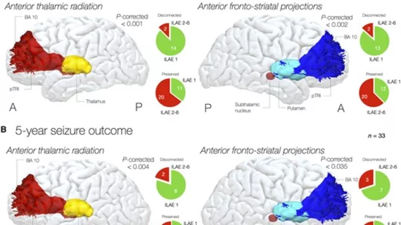
 (A) After 3 years, disconnection of anterior thalamic and striatal projections was associated with seizure freedom. Pie charts show patients being seizure free (green, ILAE 1) or seizure-relapsing (red, ILAE 2–6) according to each tract being disconnected or preserved. Each tract group includes the same tract in the left and right hemisphere. (B) Also, after 5 years, disconnection of anterior thalamic and striatal projections was associated with seizure freedom. Pie charts show patients being seizure free (green, ILAE 1) or seizure-relapsing (red, ILAE 2–6) according to each tract being disconnected or preserved.

Courtesy of Brain.

Patients who have these pathways disconnected record marked seizure-free periods of time in the long-term, according to new research.

AI company racks up 7 new FDA clearances for image triage and notification solutions

Example of data generated by an automated artificial intelligence (AI) brain CT assessment tool from Annalise.ai at RSNA 2022. What does brain imaging look like?

Example of data generated by an automated artificial intelligence (AI) brain CT assessment tool from Annalise.ai at RSNA 2022.

The Australia-based company made the announcement on April 12 in a release that described the timing of these AI-assisted solutions as “increasingly important” amid growing workloads and staffing shortages.