New DR solutions help a top New Jersey hospital lower dose, improve efficiency
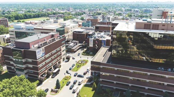
Hackensack Meridian Health Hackensack University Medical Center is the largest provider of inpatient and outpatient services in all of New Jersey. In fact, the 781-bed teaching and research hospital—which first opened its doors in Hackensack back in 1888—was ranked No. 1 in U.S. News & World Report’s 2017-2018 Best Hospital rankings for the entire state.
Topping the Garden State’s rankings, however, isn’t enough for Hackensack University Medical Center’s dedicated team of healthcare providers; the hospital is constantly bringing in new equipment to help its staff provide the absolute best patient care possible. One recent example of that ever-present drive to deliver high-quality care is the facility’s purchase and installation of a new FDR AQRO mini-digital X-ray system and five FDR Go digital radiography (DR) portable X-ray systems, all from Fujifilm.
Michael Horton, MHA, administrative director of clinical services at Hackensack University Medical Center, notes that the hospital has had a strong relationship with Fujifilm for a number of years, dating back to when it first installed the vendor’s computed radiography solutions.
“We knew based our past experience with Fujifilm, that they could deliver products built to withstand the high demand of our hospital,” Horton says. “We’re open 24/7 and have a busy emergency department—so reliability is certainly important to us. We can’t have equipment that is going to fall apart after the first two times we use it.”
Of course, Horton adds, “we also had to make sure we were buying equipment that was fiscally responsible. Fortunately, Fujifilm—as they always do—was able to offer us a beneficial price point to go along with all of the other benefits.”
Minimizing Dose a Top Priority
One of the primary reasons Hackensack University Medical Center chose Fujifilm’s DR solutions was the role they could play in helping minimize ionizing radiation across the board. Hospital employees all follow Image Gently and the ALARA (as low as reasonably achievable) principle, and it’s something they take very seriously.
“Within the imaging community, we’re all working to achieve the highest possible resolution with the lowest possible radiation dose,” Horton says. “We don’t want to expose patients to ionizing radiation when we don’t have to. We take part in dose registries to compare our doses nationally, and we work hard to keep those numbers as low as possible. If you aren’t properly watching your dose, you are doing an injustice to your patients.”
And it’s not just healthcare providers who are focused on this issue—it’s top of mind for the hospital’s patients as well.
“We always hear questions from patients, especially parents of young children undergoing an examination, about radiation,” Horton says. “It’s certainly on their minds. And it really helps that we can say to that mom or dad that, yes, we have to take an X-ray of your child, but we do have the latest and greatest equipment from Fujifilm. It helps ease their minds and reduce any concerns associated with radiation.”
Michael McGuire, M.D., a pediatric radiologist at the hospital, has noticed “a huge decrease” in radiation dose with this newer equipment, especially with pediatric and NICU patients. The radiation associated with these images was once 2-3 milliampere-seconds (mAs), he says, but now it has dropped to 0.3-0.4 mAs.
Dr. McGuire adds that this also comes into play when radiologists are faced with investigating child abuse cases for children less than a year old. “We have to do skeleton surveys for those patients,” he explains. “But in the past, we were using a pretty high dose to get the necessary details of the bones of these young kids. Now, with FDR AQRO, we decreased the dose and we get even better image quality of the bones than we ever did before. We always remember those cases. It means a lot to be able to do a better job while also using less radiation.”
Smarter Workflow, Better Images
Another benefit of installing these Fujifilm DR solutions has been a significant improvement in efficiency at Hackensack University Medical Center. The FDR AQRO, for example, is used frequently for treating pediatric and NICU patients and has greatly helped patient care in both departments.
“Workflow has greatly improved for our physicians,” McGuire says. “Our NICU doctors, for example, used to have to wait until the technologist processed each cassette before it could hit our PACS just to see the images. Now, doctors see the images right away. This has helped workflow for umbilical lines and umbilical IV lines, because the doctors can make adjustments right away as needed instead of waiting.”
Caroline Nieves, RT, chief radiologic technologist at Hackensack University Medical Center, seconds McGuire’s observation. “The imaging time is much quicker now with FDR AQRO and the FDR Go systems,” she says. “Acquiring an image used to take about three minutes, but now that time has been cut to less than half. And we can upload them so quickly to the PACS now.”
Nieves adds that the FDR AQRO “is extremely user-friendly.” Imaging NICU patients does present certain challenges, she says, but Fujifilm has built a system that is easier to use at bedside and has a much smaller footprint than Hackensack University Medical Center’s legacy equipment.
These new DR solutions are patient-friendly as well, Horton adds. At first blush, it may not seem all that helpful that the equipment is more flexible and easier to move around the hospital quietly—but that’s the kind of thing that really matters to a patient.
“Bringing in advanced technology does help relax patients,” he says. “They are going to feel better and assume they are getting better care when they see something new, sleek and quiet approaching. It is going to help them feel much more comfortable.”
As important as these benefits are to the hospital’s employees and the patients they care for around the clock, they would mean nothing if the image quality wasn’t strong. Fortunately, FDR AQRO’s images have been a hit with everyone at Hackensack University Medical Center. McGuire, for instance, says he’s seen “a big improvement in image quality, especially with pediatric and NICU patients” since the upgrade.
Nieves says she is also impressed with how good the images look when treating the hospital’s more obese patients. “You might think the images wouldn’t always come out diagnostic for bariatric patients,” she says. “But thanks to the AQRO’s Virtual Grid and new Dynamic Visualization II image processing, we can get images that look just as good as if you were using a physical grid. They look really good.”

