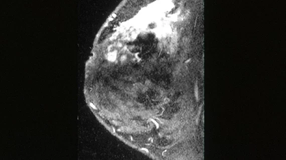
Machine learning uses MRI to predict lymphovascular invasion in breast cancer patients
Machine learning-based algorithms could help guide treatment plans for patients with breast cancer, as they may be able to accurately predict lymphovascular invasion, according to research published recently in Academic Radiology.
Tumors positive for lymphovascular invasion (LVI) may be less responsive to chemotherapy and have a poorer overall prognosis. It is, therefore, critical to understand a patient’s risk of LVI.
Typically, a postoperative biopsy is used to assess tumors for LVI but various complications can sometimes lead to misinterpretations.
“It is not always possible to detect the presence of LVI in tru-cut biopsy material; therefore, it is crucial to predict the presence of LVI from preoperative medical images,” lead author, Yasemin Kayadibi, MD, with the Department of Radiology at Istanbul University-Cerrahpasa, and co-authors explained.
Studies have shown that preoperative breast MRI findings, like prepectoral edema and adjacent vessel signs, could also hold clues that might be indicative of LVI. Researchers hypothesized that establishing LVI status at the imaging stage with the assistance of machine learning (ML) could be a promising way to direct patient treatment plans.
To test their theory, they evaluated preoperative MRIs taken from patients who had been diagnosed with invasive breast cancer between March 2015 and April 2020. In total, 128 lesions in 127 patients were included in the study.
The dataset was split into training and unseen testing sets, while 2D and 3D radiomic features were obtained from contrast-enhanced T1-weighted images and apparent diffusion-coefficient (ADC) maps.
Both the 2D and 3D models had good to excellent reproducibility, but researchers noted the 3D ADC model performed best with an accuracy of 76.7% and sensitivity of 84.6%, respectively. This model had a positive predictive value of 78.6%.
“Although studies indicate no significant difference between 2D and 3D analysis, we believe that 3D data analysis will better reflect tumor heterogeneity,” the authors explained. “Indeed, in our study, we showed that 3D analysis is superior to 2D analysis in distinguishing LVI.”
The authors go on to explain that experimenting with different techniques and machine learning-based models is crucial for future research, but they believe their study provides solid groundwork.
You can view their detailed research in Academic Radiology.

