
Deep learning reconstruction cuts breast MRI scan times in half
Deep learning reconstruction (DLR) has the potential to significantly improve the quality of breast MRIs in both accelerated and high-resolution acquisition settings.
What’s more, DLR further enhances the clinical value of breast MRI exams by shortening scan times and provides greater flexibility with protocols in comparison to conventional single-shot echo-planar imaging (ss-EPI). Researchers recently detailed the benefits of DLR in diffusion weighted breast MR settings in a new European Journal of Radiology study.
“The k-space-to-image DLR, developed based on the variational neural network is the one applied at the image reconstruction stage from k-space and has the advantage of obtaining images with good quality even with accelerated acquisition by adjusting image acquisition parameters,” Yun-Woo Chang, with the Department of Radiology at Soonchunhyang University Hospital in Seoul, Korea, and colleagues explained. “The application of DLR helps mitigate the trade-off between spatial resolution and scan time by reducing noise and compensating for signal-to-noise ratio (SNR) loss associated with high-resolution imaging.”
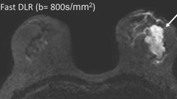
The group compared DLR exams from 50 female patients against additional images gathered via conventional techniques. These included both clinical breast MRIs and DWIs including accelerated (fast DLR) and high-resolution (HR DLR) sequences for research purposes. Two radiologists analyzed the exams for quality, which also included a comparison of ADC values to gauge performance across all three datasets.
According to the readers, both fast DLR and HR DLR significantly improved image quality in comparison to ss-EPI. The DLR protocols were consistently superior to ss-EPI for reducing artifacts across all lesion types and sizes and also resulted in a 50% reduction in scan time.
“The ability to reduce scan time by approximately 50% while improving image quality presents significant benefits, streamlining workflow without compromising diagnostic accuracy and reducing patient discomfort," the authors wrote. "In addition, the versatility of DLR in high-resolution acquisition setting allows for tailored parameter adjustments to meet clinical demands, further enhancing its applicability.”
ADC values for fast DLR also showed lower standard deviation compared to ss-EPI in malignant, mass-type lesions and in lesions smaller than 2 cm. This is likely owed to the reduced artifacts and noise afforded by the DLR protocols, the group noted.
The authors suggested their findings hold “significant clinical relevance,” though additional research is needed to further assess the value of DLR protocols in independent screening settings.
Learn more about the findings here.

In addition to her background in journalism, Hannah also has patient-facing experience in clinical settings, having spent more than 12 years working as a registered rad tech. She began covering the medical imaging industry for Innovate Healthcare in 2021.