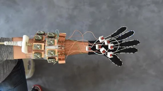
'Glove' prototype captures MRI of joints in action
MRI, for all it can do, has many design limitations—but an NYU School of Medicine team recently created an MRI device that users can slip on like a glove to produce high quality images of moving joints.
"Our results represent the first demonstration of an MRI technology that is both flexible and sensitive enough to capture the complexity of soft-tissue mechanics in the hand," said lead author Bei Zhang, PhD, research scientist at the Center for Advanced Imaging Innovation and Research (CAI2R), within the Department of Radiology at NYU Langone Health, in a release.
The group’s glove prototype can help diagnose strain injuries, guide surgery with hand images, and aid in designing prosthetics, according to authors of the study published online May 4 in Nature Biomedical Engineering.
While most MRIs utilize “low impedance” receiver coils allowing current to flow easily, this new device uses high impedance to block current and measure how hard the force in magnetic waves “push” when it attempts to establish a current.
With no current involved, the coils no longer require a rigid structure and researchers say their new flexible coil system produces high-quality images of freely moving muscles, tendons and ligaments in the hand.
"We wanted to try our new elements in an application that could never be done with traditional coils, and settled on an attempt to capture images with a glove," said senior author Martijn Cloos, PhD, assistant professor from the CAI2R institute in the department of radiology at NYU Langone Health, in the release. "We hope that this result ushers in a new era of MRI design, perhaps including flexible sleeve arrays around injured knees, or comfy beanies to study the developing brains of newborns."

