MRI-compatible ultrasound system may improve flexibility
A new ultrasound prototype utilizing an optical—rather than electronic—system may provide flexibility in simultaneously pairing the modality with other imaging techniques, such as MRI. Experts say it could provide doctors more options for using ultrasound to diagnose and treat patients.
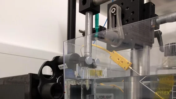
Led by Erwin J. Alles with University College London, researchers created the all-optical ultrasound system that uses light beam scanning mirrors to create video-rate, real-time 2D images. Users can toggle between various modes to capture different ultrasound image types, which is typically done with separate specialized probes, according to a news release from the Optical Society.
“All-optical ultrasound imaging probes have the potential to revolutionize image-guided interventions,” Alles said in the release. “A lack of electronics and the resulting MRI compatibility will allow for true multimodality image guidance, with probes that are potentially just a fraction of the cost of conventional electronic counterparts.”
The team tested their machine on a dead zebrafish and a pig artery which was manipulated to mimic the dynamics of pulsing blood. Results published in Biomedical Optics Express showed imaging capabilities comparable to an electronic high-frequency ultrasound system. It achieved a sustained frame rate of 15 hertz, a dynamic range of 30 decibels, a six-millimeter penetration depth and a 75 by 100 micrometer resolution.
Previously, the team used all-optical ultrasound to create 2D and 3D images, but the method took hours. The new system is faster and clearer, researchers noted.
“Through the combination of a new imaging paradigm, new optical ultrasound generating materials, optimized ultrasound source geometries and a highly sensitive fiber-optic ultrasound detector, we achieved image frame rates that were up to three orders of magnitude faster than the current state-of-the-art,” Alles said.

