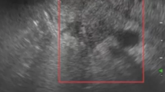
Multimodal AI model helps differentiate between benign and malignant pancreatic lesions
Artificial intelligence can improve the performance of novice endoscopists conducting ultrasound procedures on solid pancreatic lesions, improving clinical diagnoses.
Endoscopic ultrasonography (EUS) has emerged as a valuable tool for diagnosing pancreatic cancer, but its specificity in differentiating between benign and malignant pancreatic lesions is lacking. Further, operator experience has a significant impact on performance, creating questions about the modality’s diagnostic accuracy, authors of a new analysis in JAMA Network Open explain.
“Given the deep learning curve of the EUS examination and the lack of standardized and sufficient training procedures, the diagnostic ability varies greatly among endoscopists, particularly for less-experienced individuals,” Aiming Yang, MD, from the department of gastroenterology at Peking Union Medical College Hospital in China, and co-authors note. “Artificial intelligence has the potential to help, but existing AI models focus solely on a single modality.”
For the study, the team developed and deployed a multimodal AI model that combines imaging and clinical information to reach a diagnosis. Twelve endoscopists with varying experience levels tested the model on datasets of over 600 patients with solid pancreatic lesions. The endoscopists were randomly assigned to two groups—one that utilized AI assistance and one that did not.
The AUC of the joint AI model ranged from 0.996 in the internal test dataset to 0.955, 0.924, and 0.976 in three external test datasets, respectively. The model significantly improved the performance of endoscopists with less experience, while those who were more experienced reported higher confidence using AI.
These findings indicate that the multimodal model could help address the learning curve associated with EUS, the authors suggest.
“The interaction between endoscopists and the joint-AI model can potentially ameliorate this situation, as the performance of novice endoscopists was significantly improved with the AI assistance in the crossover trial,” the group writes. “The transparency of the decision-making process in medical practice is highly valued.”
The model also provided readers with an interpretability analysis that helped them understand how it arrived at a specific diagnosis. This increased trust among the more experienced endoscopists.
This sort of model transparency is key when it comes to AI, the group notes, adding that transparency should be a central focus in future AI research.
The full study can be accessed here.

