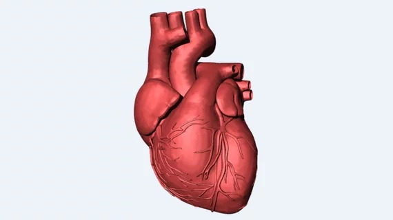
3D printing workflow utilizes CT scans to improve aortic valve replacement
With the development of a new three-dimensional (3D) printing workflow, researchers at the Wyss Institute for Biologically Inspired Engineering at Harvard University in Boston may help cardiologists produce more proficient implantable heart valves and determine how different sized valves interact for each patient before surgical intervention for aortic valve replacement, according to a Dec. 10 report by the 3D Printing Media Network.
The workflow uses CT scans of the patient’s aorta to produce virtual 3D models that are then printed into physical models. Additionally, a “sizer” device designed to fit inside the 3D printed valve models is also printed, according to the story.
The sizer—which has its own unique shape and amount of calcification—can expand and contract to determine an appropriately sized transcatheter aortic valve replacement (TAVR), according to the article.
The researchers hope their 3D printing workflow can help cardiologists see “leaflets” of tissue that open and close inside the aorta, which are too thin to be seen on a standard CT scan.
“In sizing replacement TAVR valves, doctors don’t get the opportunity to evaluate how a specific valve size will fit with a patient’s anatomy before surgery,” study author James Weaver, PhD, senior research scientist at the Wyss Institute, told the news outlet. “Our integrative 3D printing and valve sizing system provide a customized report of every patient’s unique aortic valve shape, removing a lot of the guesswork and helping each patient receive a more accurately sized valve.”
See the entire article below.

