Augmented reality helps cardiologists visualize 3D images, plan complex procedures
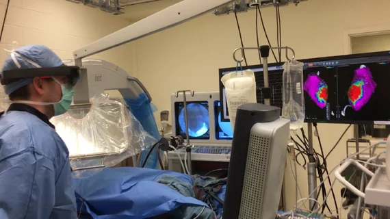
Augmented reality (AR) allows cardiologists to visualize and explore three-dimensional images of myocardial scarring in the heart as they perform ventricular tachycardia ablation or other electrophysiological surgical interventions, according to research published online Oct. 8 in PLOS One.
The AR technology allows physicians to superimpose MRI or CT scans to help guide therapeutic interventions and give surgeons the opportunity to interact with medical data without physically touching a screen. It's goal is to maintain a sterile environment and reduce the risk of infection.
“Our report shows exciting potential that having this complex 3D scar information through augmented reality during the intervention may help guide treatment and ultimately improve patient care,” author Jihye Jang, a PhD candidate at the Cardiac Magnetic Resonance (MR) Center at Beth Israel Deaconess Medical Center in Boston, said in a prepared statement.
The researchers utilized Microsoft’s HoloLens to generate 3D holographic myocardial scar tissue from five animal models that underwent controlled infarction and electrophysiological study.
Additionally, an operator and mapping specialist analyzed the holographic 3D scarring during electrophysiological study and completed a questionnaire regarding the feasability of combining holographic 3D late gadolinium enhancement data with real-world environments, such as a surgical suite or an actual patient.
“Our report is one of the first efforts to test augmented reality in cardiovascular electrophysiological intervention,” Jang said. “Our next steps will expand the use of AR into treatments for arrhythmia by merging the scar information with electrophysiology data.”
The National Institutes of Health and the American Heart Association supported the research.

