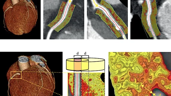
Novel cardiac imaging biomarker could predict risk of coronary inflammation, heart attack
A team of international researchers has developed a new imaging biomarker able to non-invasively predict a patient’s risk of coronary inflammation and heart attack, according to research published Aug. 28 in The Lancet.
Experts from Cleveland Clinic, the University of Oxford and the University of Erlangen in Germany created the biomarker to measure inflammation of fatty tissue surrounding the coronary arteries.
“This is an exciting new technology which has the potential for providing a simple, non-invasive answer to detect patients at risk for future fatal heart attacks," said co-author Milind Desai, MD, a cardiologist at Cleveland Clinic, in a prepared statement. “More importantly, it highlights the incredible value of cross-continent collaboration to validate the findings in different populations.”
Desai and colleagues developed the perivascular fat attenuation index (FAI) as an imaging biomarker, which can map changes in perivascular fat levels on coronary computed tomography angiography (CTA) scans and may improve detection of inflammation.
For the study, called Cardiovascular Risk Prediction using Computed Tomography (CRISP-CT), researchers recruited two cohorts of patients undergoing CTA scans—1,872 patients (average age of 62 years) in Germany from 2005 to 2009 and 2,040 patients (53 years) at Cleveland Clinic from 2008 to 2016.
Higher perivascular FAI values in both cohorts indicated greater coronary inflammation and were associated with high rates of cardiac mortality, according to the researchers.
Overall, the new biomarker may improve primary and secondary prevention of cardiovascular risk, explained lead author Charalambos Antoniades, MD, PhD, of the University of Oxford, who believes the biomarkers could become an integral part of CTA.
“For the first time we have a set of biomarkers, derived from a routine test that is already used in everyday clinical practice, that measures what we call the ‘residual cardiovascular risk’, currently missed by all risk scores and non-invasive tests,” said Antoniades, who led the study at the University of Oxford’s division of cardiovascular medicine.
The research was presented this week at the European Society of Cardiology (ESC) Congress 2018 in Munich.

