Is MRI better than angiography for diagnosing, treating certain heart attacks?
Cardiac magnetic resonance imaging (MRI) may better assess and treat patients who have experienced a type of heart attack caused by an extremely narrowed artery compared to standard angiography, reported authors of a May study published in Circulation.
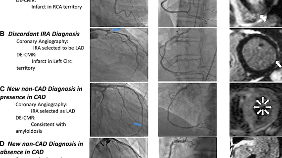
Overall, the use of cardiac MRI led to a new diagnosis involving a damaged artery in 30% of patients, and a diagnosis unrelated to the artery in another 15%, reported John Heitner, MD, director of noninvasive imaging at NewYork-Presbyterian Brooklyn Methodist Hospital, and colleagues.
Heitner et al. explained that it can be hard for physicians to identify the damaged artery responsible for a non-ST segment elevation myocardial infarction (NSTEMI). Typically their first choice is coronary angiography, with MRI only considered afterward and if results remain unclear.
“We sought to learn whether an MRI performed first could improve clinicians’ ability to identify the affected artery,” Heitner said in a prepared statement. “Many patients with NSTEMI have diseases in several blood vessels or have no significant blockages at all that make it difficult to identify the correct artery that caused the heart attack. We suspected the MRI's ability to directly visualize the heart muscle that was affected could improve accuracy, and our research suggests that to be the case."
One-hundred and fourteen patients were enrolled in the study, all had presented with their first heart attack at one of three centers. Everyone underwent an MRI and subsequent angiography; images were independently and blindly reviewed to locate the clogged artery responsible for the heart attack.
Readers couldn’t identify the blocked artery using angiography in 37% of patients. Of that group, MRI successfully located the artery in 60% of patients, or led to a new non-coronary artery disease diagnosis in 19%.
Among cases when angiography successfully found the blocked artery, MRI found a second artery in 14% and a non-CAD diagnosis in another 13%.
“This study suggests that using an MRI should be the standard practice in NSTEMI patient diagnosis and care,” Heitner said. “We want to make sure that we are treating the artery that caused the heart attack as this will likely lead to better long term outcomes for the patient."

