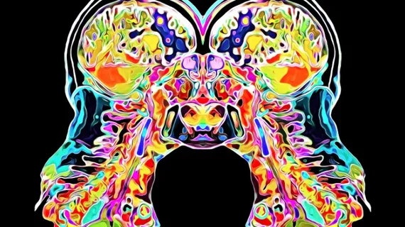
Toronto physician creates anatomical art from medical images
When not administering therapy to patients who had heart surgery, Trinley Dorje, a physiotherapy assistant at Peter Munk Cardiac Center in Toronto, is creating anatomical art from medical images of the human body.
Melding her passions for anatomy and art, Dorje’s work offers an aesthetically pleasing and anatomically correct perspective into how the human body functions that could otherwise only be seen at a hospital, according to a report published Aug. 15 by CBC in Canada.
"That's why I really decided to use medical imaging as a form of focus for my art, because it's a way that people don't normally see their anatomy," Dorje told CBC.
Although remaining anatomically correct while illustrating medical images is a challenge with every piece she creates, Dorje explained that creating her work helps her manage her emotions from what she sees at the hospital, according to the article.
"I think it's almost a therapy for me to be able to create these images which display sometimes which would be catastrophic events even, like strokes and things like that within an artwork,” Dorje said. “But at the same time, I'm making them as pretty as I can."
See the CBC’s entire article below.

