Severe COVID patients continue to evidence lung abnormalities 2 years after initial infection
Two-year follow-up data from patients who had severe COVID reveals that some continue to display lung abnormalities two years after initially acquiring the virus [1].
These latest findings were published Feb. 14 in Radiology.
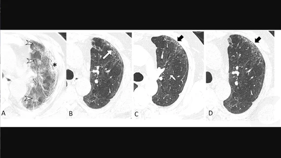
The analysis includes chest CT scans from a cohort of 144 patients who had been hospitalized with COVID between January 15 and March 10, 2020. Each participant underwent a total of three chest CTs in order to be included in the analysis—one at six months, 12 months and two years following the onset of their symptoms.
Those scans showed a combination of fibrosis, thickening, honeycombing, cystic changes and dilation of the bronchi, among other imaging features.
Co-senior authors of the study Qing Ye, MD, and Heshui Shi, MD, PhD, both with Tongji Medical College of Huazhong University of Science and Technology in Wuhan, China, and colleagues noted that the finding of fibrosis is of particular concern.
“In particular, the proportion of fibrotic interstitial lung abnormalities, an important precursor to idiopathic pulmonary fibrosis, remained stable throughout follow-up,” the authors said. “Therefore, the fibrotic abnormalities observed in our study might represent a stable, irreversible pulmonary condition, such as lung fibrosis, after COVID-19.”
The good news is that many of the other lung abnormalities visualized at six months appeared to decrease over time. At six months, 54% of patients showed abnormalities, but by the two-year mark, that figure decreased to 39%, with 61% showing complete radiological resolution.
Of the patients who continued to show abnormalities on imaging, 23% were found to be fibrotic, and 22% still harbored respiratory symptoms and decreased lung function. For the symptomatic, the most common issue was noted as exertional dyspnea or shortness of breath. The authors suggested that these lingering symptoms could likely be attributed to their ongoing lung damage, but that the “long-term and functional consequences of chest CT findings post-COVID-19 are largely unknown.”
The experts suggested that patients who continue to show respiratory symptoms following the recovery from their initial infection need to be continuously monitored for functional impairment.
The study abstract is available here.

