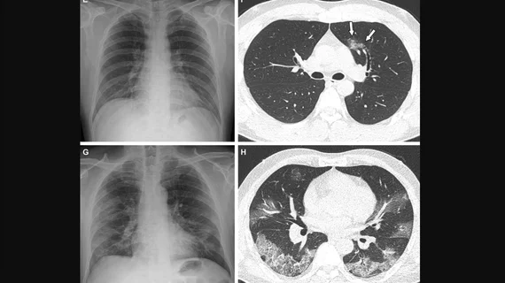
Vaccinated vs unvaccinated, Delta vs Omicron—how do these factors impact clinical and imaging features?
Research has established differing characteristics of each COVID variant and how vaccination status impacts clinical outcomes, but how do these varying factors differentiate on imaging?
A new paper published in Radiology presents clinical and imaging data from more than 2,000 patients to lay the groundwork for answering this question. Experts focused their attention on how the Omicron and Delta variants, in addition to vaccination status, affect disease severity and imaging—an area of COVID research that has not yet been thoroughly explored, according to the authors of the study.
“Several recently published studies revealed that clinical course and pneumonia severity are somewhat different according to the viral variants,” corresponding author Yeon Joo Jeong, MD, PhD, of the Research Institute for Convergence of Biomedical Science and Technology at Pusan National University Yangsan Hospital, and colleagues explained. “However, few reports have evaluated the effect of both vaccination and viral variant prevalence on clinical and imaging severity of COVID-19.”
The authors found that patients who were fully vaccinated fared the best in terms of clinical severity [1]. Based on imaging, these patients also had the fewest cases of severe pneumonia and were more likely to have negative chest radiographs and CT scans.
A comparison between COVID variants (1,022 Delta and 1,158 Omicron) revealed that patients who were diagnosed with Delta were more likely to display severe pneumonia on CT and had worse overall clinical outcomes (based on ICU admission and in-hospital deaths).
Vaccination rates were also found to be lower during the Delta wave, the authors shared, adding that this could play a large role in why disease and pneumonia severity were both found to be reduced during the wave of the Omicron variant. The results of this study further support the notion that being fully vaccinated against COVID has numerous protective benefits, the authors suggested.
For more insight from the research, click here.

